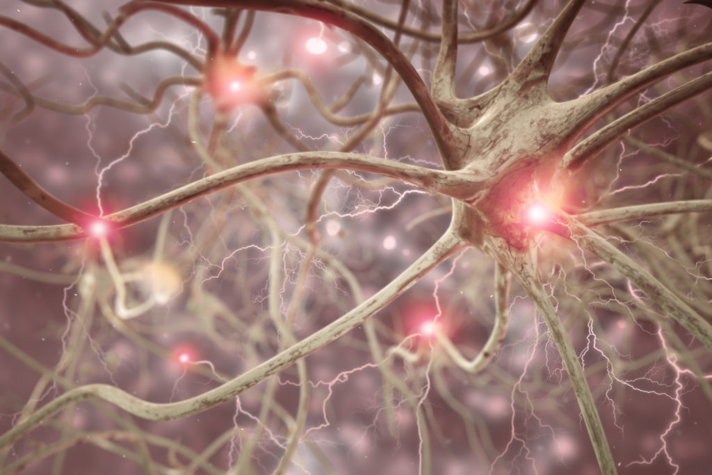
Researchers have recently identified a neuron which is crucial for navigation in mice’s brains.

Scientists at Ann Arbor University of Michigan have found a previously unrecognized excitatory neuron in the brains of the mouse. The neuron is key to navigation, they claim.
This discovery, now published in the Cell Reports journal, can help scientists understand how the part of the brain that is responsible for navigation— the retrosplenial cortex (RSC) — goes about navigating long distances.
Navigation and neurology
Scientists know that navigation is important to the RSC. In fact, if a person’s RSC is compromised, they will not only experience memory loss but also greatly reduce their ability to navigate.
A person with a damaged RSC, for instance, can find it very difficult to navigate their usual route from work to home.
Scientists, however, are less specific about how the parts of this cortex interact, so a person can navigate successfully.
The researchers gained information about what this new neuron does, as well as what the other key neuron in the RSC does, by identifying this previously unrecognized excitatory neuron.
Two types of neuron
The researchers studied the mice’s RSCs and produced computational models capable of simulating the realistic reactions of RSC cells to different types of information.
This allowed them to determine that there were two separate neurons focused on different forms of navigational information.
One form, called common regular-spiking (RS) neurons, was responding to head direction changes. The other type, called the neurons with low rheobase (LR), responded to the head maintaining a consistent direction.
That’s what the neurons do because of variations in how they get information.
The RS neurons respond to significant changes but are unable to continue firing to receive more information after this. This is because after the initial significant change in direction their signals are getting slower.
The LR neurons, by contrast, fire consistently and faster, and can respond to subtler directional changes. This means they can detect very small variations in the head position which suggest a continuous direction of motion.
‘The Little Neuron That Could’
According to study co-author Ellen Brennan, who named the LR neurons, “A better name for this tiny yet tenacious little neuron, as proposed by my classmate, would be’ The Little Neuron That Could.'”
“It’s the perfect name because it illustrates the resilience that makes them optimally adapted to the continued path of code.
As the lead author of the study Dr. Omar Ahmed explains: “Regular neurons in the cortex are only good at encoding directional information when you move your head, but what happens if your head is still there? You still need to know what direction you’re facing so you can plan your route using this information.
” Ideally, you need another kind of neuron— a neuron that can encode your orientation continuously over long durations even when your head is not moving.
The RSC needs those two neurons to work together to help navigate. As Brennan states in a video detailing the results, it is similar to having a compass but not understanding how North is not having LR neurons that sense directional movement.
A link to Alzheimer’s disease?
The researchers are now researching whether or not these results would be useful in understanding how Alzheimer’s disease affects those neurons. The most common type of dementia is in Alzheimer’s. It affects the spatial awareness of a individual, inter alia. There is actually no treatment for that disease.
As Dr. Ahmed notes: “The retrosplenial cortex is important for spatial orientation but it is one of the first brain regions in[ people with Alzheimer’s] to display impaired behavior. That is probably why the vast majority of[ people with the history of Alzheimer’s] spatial disorientation can easily get confused — because their retrosplenial cells are not functioning as they should.
“By understanding how retrosplenial cells encode compass-like information in healthy versus Alzheimer’s brains, we hope to start working toward novel therapies.”
– Dr. Omar Ahmed


