Electromyography Device: Portable, Low-Noise EMG/NCS Platform

All Categories

Electromyography Device for Professional EMG and Nerve Conduction Diagnostics

An Electromyography Device is a specialized neurophysiology system used to record, analyze and document muscle and peripheral nerve activity for clinical diagnosis and research. A modern Electromyography Device combines portable hardware, low‑noise amplifiers and intelligent analysis software so that clinicians can perform needle EMG, surface EMG and nerve conduction studies efficiently in outpatient clinics, inpatient wards and rehabilitation centers. With options for wireless data transmission and bedside operation, the Electromyography Device improves patient comfort, especially for individuals with limited mobility, and supports point‑of‑care decision‑making. For hospitals, laboratories and distributors, an Electromyography Device is not only a testing instrument but also a standardized data and reporting platform that enhances workflow, supports multi‑modal neurophysiology services and integrates easily with hospital information systems.
Get A Quote

Electromyography Device

A high‑quality Electromyography Device is central to any neuro‑diagnostic laboratory, because it determines both diagnostic accuracy and patient experience. By combining portable design, precise signal acquisition and intelligent software, an Electromyography Device helps neurologists and physiatrists complete complex studies with greater confidence and efficiency. For B2B customers, a scalable Electromyography Device platform also creates recurring value through consumables, software upgrades, service contracts and research collaborations.

Portable and Wireless Electromyography Device for Flexible Use

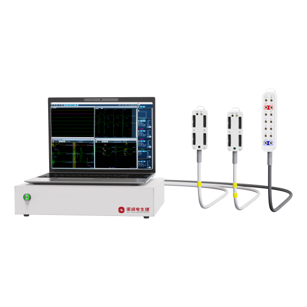
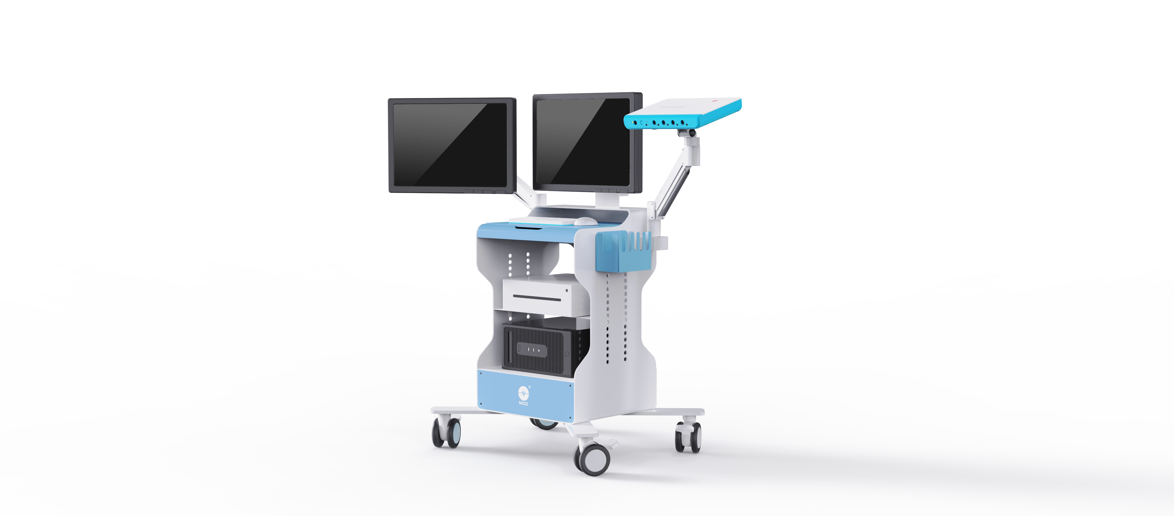
A modern Electromyography Device is designed to be compact, lightweight and easy to move, replacing the traditional bulky workstation that must stay in a shielded room. With trolley, tabletop or handheld configurations and wireless or Bluetooth data links, an Electromyography Device can be brought directly to the bedside, ICU or rehabilitation gym. This flexibility allows clinicians to examine patients who cannot easily travel to a dedicated lab and enables sports medicine teams or researchers to deploy the Electromyography Device in field environments. For hospitals and distributors, the same Electromyography Device can support multiple departments, improving utilization rates and making capital investment more efficient.

High-Precision, Low-Noise Signal Acquisition in the Electromyography Device

The core technical value of an Electromyography Device lies in its ability to capture very small EMG signals with high fidelity and minimal noise. Needle EMG and surface EMG recordings are typically in the microvolt range and are easily contaminated by mains interference, motion artifacts and electromagnetic noise from other devices. A premium Electromyography Device uses high‑grade differential amplifiers with excellent common‑mode rejection, high‑resolution analog‑to‑digital conversion and carefully shielded cables to preserve waveform integrity. As a result, the Electromyography Device can clearly display motor unit potentials, insertional activity and recruitment patterns, supporting accurate distinction between neurogenic and myopathic disorders.

Intelligent Software and Multi-Modal Integration in One Electromyography Device

Today’s Electromyography Device is not just a simple recorder; it is an intelligent diagnostic platform. Built‑in software offers automatic artifact detection, motor unit potential decomposition, interference pattern analysis and automated calculation of latency, amplitude and conduction velocity. A single Electromyography Device can integrate nerve conduction studies, needle EMG, F‑wave, H‑reflex and repetitive nerve stimulation, allowing comprehensive evaluation of peripheral nerves, muscles and neuromuscular junction within one system. Structured databases and standardized report templates enable the Electromyography Device to generate clear clinical reports quickly, reducing manual documentation work and supporting research and teaching.

Related Products

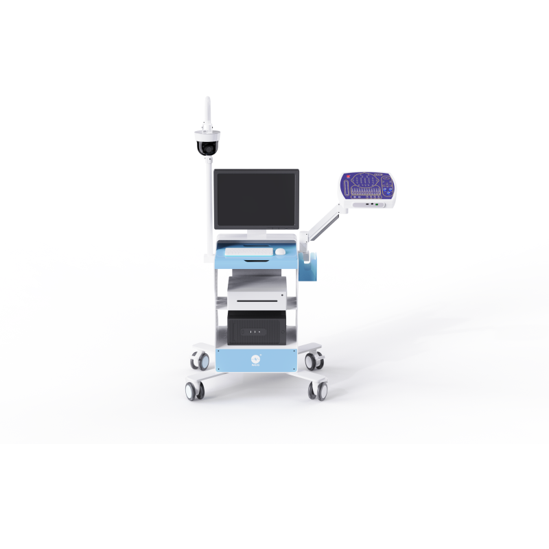
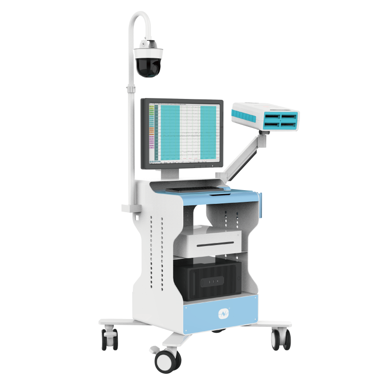
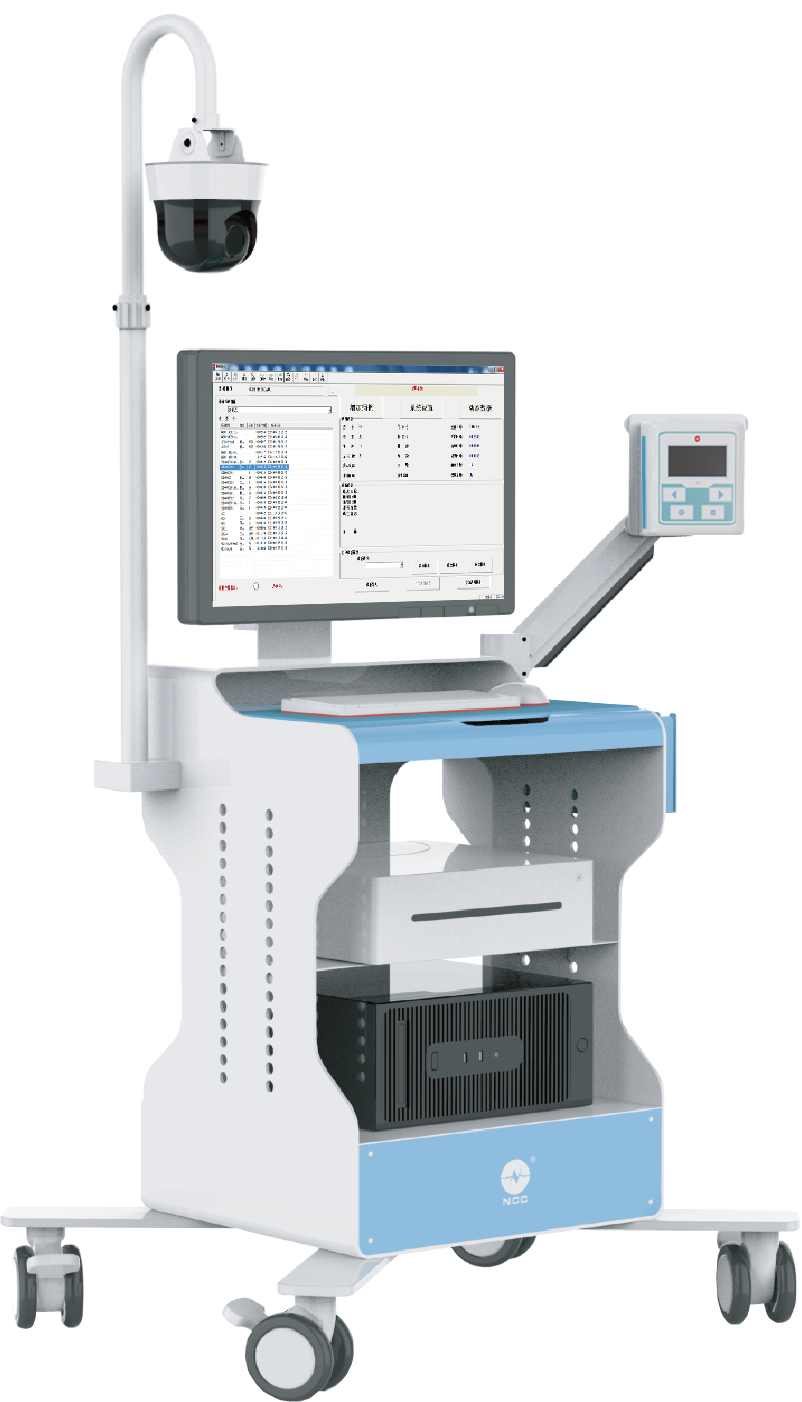
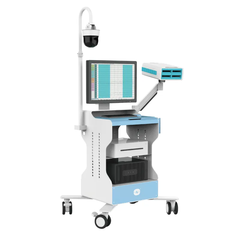
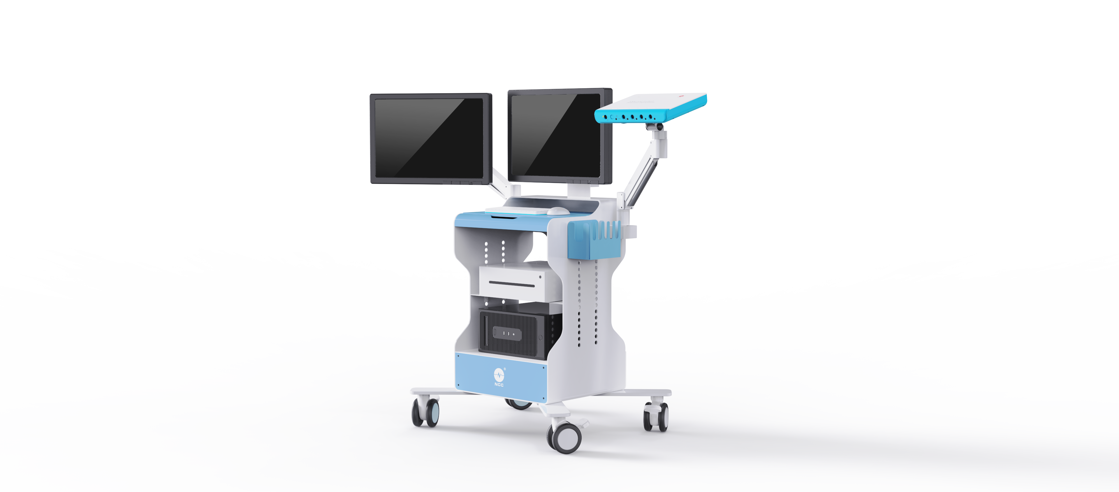
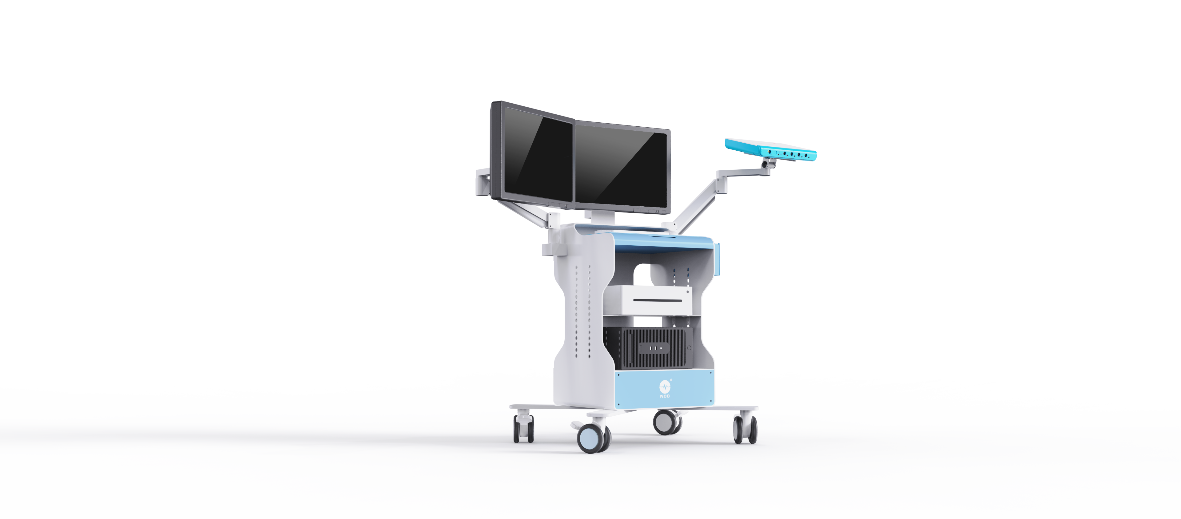
Our Electromyography Device is a high‑performance neurophysiology platform engineered to support routine and advanced EMG and nerve conduction diagnostics. The hardware unit of the Electromyography Device integrates high‑input‑impedance amplifiers, wide dynamic range and low intrinsic noise, ensuring clear recording of both needle EMG and surface EMG signals across the required frequency spectrum. Carefully designed shielding, optimized grounding and high‑quality cables reduce interference from mains power and other medical equipment. The Electromyography Device can be configured as a compact cart‑based station or as a portable module connected to a laptop or tablet, depending on the clinical environment. On the software side, user‑friendly workflows guide clinicians through standardized nerve conduction and EMG protocols, with automated measurements, reference comparisons and customizable templates.

As a specialized neuro‑diagnostic and neuromonitoring manufacturer, NCC Medical Group develops and produces complete Electromyography Device solutions for hospitals, clinics and research institutions worldwide. Established in 1997, NCC has grown from a small electrophysiology laboratory into a recognized provider of advanced IONM, EEG/PSG, EMG and medical consumables. Our Electromyography Device portfolio reflects this long‑term focus on neurophysiology: we offer systems that range from entry‑level EMG workstations for community hospitals to high‑channel, multi‑modal Electromyography Device platforms for academic centers and research labs.

NCC’s R&D and manufacturing operations are based in Shanghai, where dedicated engineering teams design the electronics, software and mechanics of each Electromyography Device. We operate modern production lines that include automated SMT assembly, in‑circuit testing and final system calibration. Every Electromyography Device undergoes a defined quality control process, including noise performance checks, signal linearity tests and long‑duration stability verification. This ensures that the Electromyography Device performs consistently even in demanding clinical environments. Our quality and compliance hub in Singapore coordinates international certifications and regulatory submissions, making sure that each Electromyography Device meets relevant standards in Asia, Europe, the Americas and other regions.

To support seamless global operations, NCC structures its business around three pillars. The R&D and manufacturing pillar focuses on core technology for the Electromyography Device, including amplifier design, artifact rejection algorithms and intelligent analysis modules. The global sales and export pillar manages distribution channels, OEM/ODM partnerships and regional marketing, helping local partners position the Electromyography Device effectively in their markets. The service and innovation pillar operates the NeuGuard Service Platform and the NCC Innovation Academy, providing training, remote technical support and continuous education related to the Electromyography Device and other neurophysiology products.

NCC’s solutions, including the Electromyography Device, are deployed in thousands of hospitals in China and more than 50 countries worldwide. Our customers range from top‑tier teaching hospitals that use the Electromyography Device for complex neuro‑muscular diagnostics and research, to regional centers that rely on the system for routine nerve conduction and EMG studies. We maintain 31 training and technical support centers across China, where clinicians and technicians receive hands‑on instruction in Electromyography Device operation, troubleshooting and interpretation. Internationally, we are actively expanding a distributor network and offer online training, webinars and on‑site startup support to ensure that each Electromyography Device is integrated smoothly into local workflows.

As a National “Little Giant” (SRDI) enterprise, NCC is recognized for Specialized, Refined, Differential and Innovational capabilities. This designation reflects our commitment to continuous innovation in areas such as intelligent EMG analysis, wireless acquisition modules and integrated multi‑modal platforms. Our mission is to safeguard life and health by providing tools that support safer surgeries, precise diagnostics and high‑quality patient care. The Electromyography Device is a key element of this mission, enabling clinicians to detect neuromuscular disorders earlier and manage them more effectively. For B2B partners, NCC offers flexible collaboration models around the Electromyography Device, including branding customization, interface localization and application‑specific software options, ensuring that each market can build a tailored neurophysiology solution on top of a robust, proven technology base.

FAQ

What examinations can an Electromyography Device perform?

An Electromyography Device can perform a broad range of neurophysiological examinations that are essential for diagnosing neuromuscular disorders. The core functions of an Electromyography Device include nerve conduction studies to evaluate peripheral nerve conduction velocity and latency, needle EMG to assess motor unit potentials and muscle recruitment, and surface EMG for non‑invasive muscle activity recording. In addition, an Electromyography Device typically supports F‑wave and H‑reflex tests to evaluate proximal nerve segments and spinal reflex arcs, as well as repetitive nerve stimulation for neuromuscular junction assessment in conditions such as myasthenia gravis. By combining these techniques, the Electromyography Device allows clinicians to examine the entire peripheral nervous system from nerve roots to neuromuscular junction and muscle.
An Electromyography Device improves diagnostic accuracy by providing high‑quality signals and advanced analysis tools that reveal subtle abnormalities in nerve and muscle function. The use of low‑noise amplifiers and high‑resolution converters ensures that the Electromyography Device can display small motor unit potentials clearly, which is crucial when differentiating neurogenic and myopathic patterns. Intelligent software functions such as automatic measurement of latency, amplitude and duration, motor unit potential decomposition and interference pattern analysis help standardize examinations and reduce operator‑dependent variability. In addition, the Electromyography Device allows comparison of measured values with reference data stored in the system, supporting evidence‑based interpretation. Together, these features allow clinicians to use the Electromyography Device to reach more precise and reproducible conclusions.
To implement an Electromyography Device, a hospital needs a suitable physical space, basic power and network connections and trained personnel. The examination room should provide a reasonably low‑noise environment and comfortable conditions for patients undergoing needle EMG and nerve conduction tests. Technicians and physicians must be trained in electrode placement, stimulation parameter selection and interpretation of findings produced by the Electromyography Device. From an IT perspective, integration with hospital information systems or PACS can be arranged so that reports and selected waveforms from the Electromyography Device are stored centrally. NCC and its partners support customers with installation, on‑site or online training, protocol templates and ongoing technical service to ensure that the Electromyography Device becomes a reliable part of the daily diagnostic workflow.

More Posts

Market Dynamics of Portable EEG Price and Procurement Cost Optimization Solutions

04

Mar

Market Dynamics of Portable EEG Price and Procurement Cost Optimization Solutions

Introduction: Reframing the Meaning of Portable EEG Price in 2026 In 2025, the global portable EEG market reached an estimated value of USD 92 million and is projected to grow to approximately USD 153 million by 2031, representing a compound annual g...
View More
Technical Advantages and Stable Supply Analysis of Professional EMG Stimulators in Clinical Diagnostics

04

Mar

Technical Advantages and Stable Supply Analysis of Professional EMG Stimulators in Clinical Diagnostics

In clinical neurophysiology, the emg stimulator is often underestimated. It appears, at first glance, to be a controlled current source—an accessory to the electromyography system. In reality, it is the metrological reference upon which n...
View More
Clinical Advantages and Supply Chain Assurance of Wireless EEG Devices

05

Mar

Clinical Advantages and Supply Chain Assurance of Wireless EEG Devices

Wireless EEG device technology is redefining the operational boundaries of neurodiagnostics. What was once confined to fixed electroencephalography laboratories is now extending into intensive care units, emergency departments, ambulatory environment...
View More
Compliance and Technical Advantages of Portable EEG Machines: Empowering Efficient Clinical Neuro Monitoring in Healthcare Institutions

05

Mar

Compliance and Technical Advantages of Portable EEG Machines: Empowering Efficient Clinical Neuro Monitoring in Healthcare Institutions

The true value of an eeg machine portable solution is not measured by how small it is, but by how reliably it performs when removed from the controlled environment of a neurophysiology laboratory and placed into the complexity of real clinical care. ...
View More

Customer Reviews

Dr. Emily Carter, Neurologist

“Introducing the new Electromyography Device into our neurophysiology lab has significantly improved both workflow and diagnostic confidence. The portable design allows us to move the Electromyography Device between outpatient clinic and ward without difficulty, and the low‑noise amplifiers provide very clear needle EMG signals, even in challenging cases. Automated nerve conduction measurements and reference value comparisons save time and make it easier for our junior staff to learn. Overall, the Electromyography Device has helped us standardize examinations and deliver more consistent reports to referring physicians.”

Dr. Luca Bianchi, Physiatrist

“As a rehabilitation specialist, I use the Electromyography Device not only for diagnosis but also to track progress in neuromuscular recovery. The system’s ability to perform both surface EMG and needle EMG, combined with nerve conduction studies, gives me a comprehensive view of each patient’s status. The user interface of the Electromyography Device is intuitive, and I can quickly access previous sessions for comparison. This has made it easier to adjust therapy plans based on objective data from the Electromyography Device rather than relying only on subjective reports and manual muscle testing.”

Ms. Sarah Novak, Clinical Neurophysiology Lab Manager

“From a management perspective, the Electromyography Device has been a very positive investment for our hospital. Standardized templates and structured reporting have reduced dictation time and improved the consistency of our EMG reports. The database within the Electromyography Device also makes it simple to search past cases for teaching or quality audits. Our technicians appreciate the ergonomic design and the reliability of the Electromyography Device during long testing days. With responsive support from the manufacturer, we feel confident using this platform as the backbone of our neurodiagnostic service.”

Get a Free Quote

Our representative will contact you soon.
Email
Name
Company Name
Message
0/1000
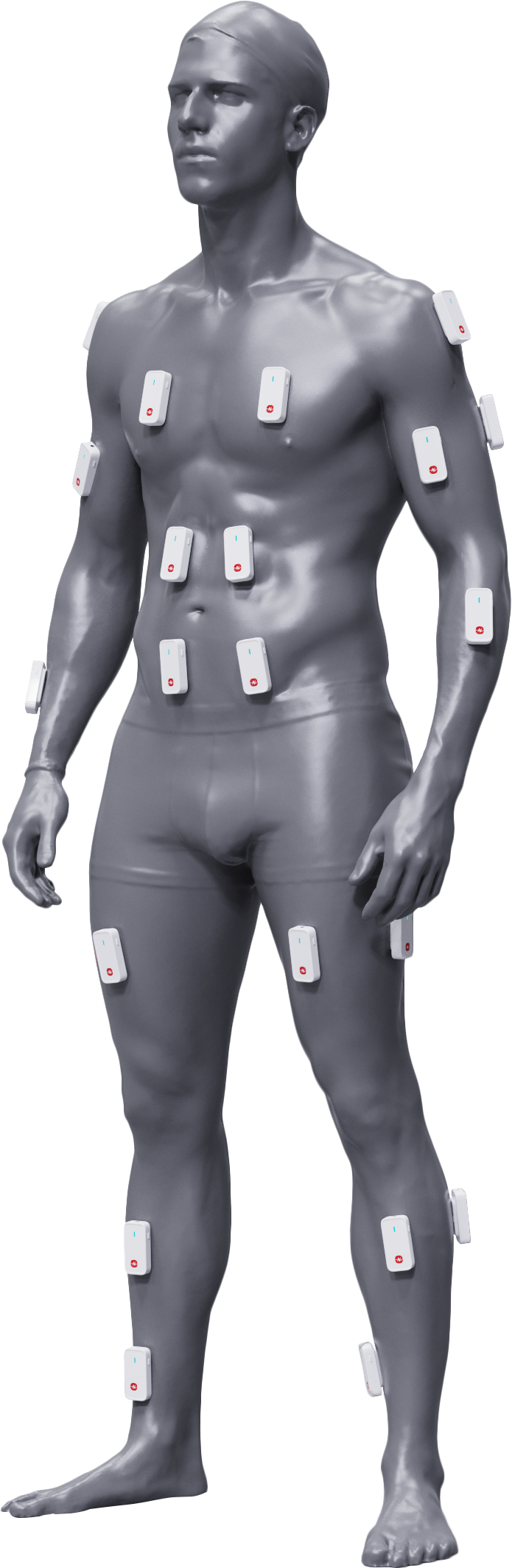
Portable Electromyography Device for Bedside and Field Use

Portable Electromyography Device for Bedside and Field Use

The Electromyography Device features a compact, lightweight design with optional wireless connectivity, allowing clinicians to perform EMG and nerve conduction tests directly at the bedside, in the ICU or in rehabilitation and sports settings. This mobility increases patient comfort and extends the reach of neurophysiology services.
High-Fidelity EMG Signals from the Electromyography Device

High-Fidelity EMG Signals from the Electromyography Device

With high‑performance differential amplifiers, excellent common‑mode rejection and robust shielding, the Electromyography Device delivers clean, detailed EMG signals even in electrically noisy clinical environments. This high fidelity supports accurate interpretation of motor unit potential morphology and recruitment patterns.
Integrated Multi-Modal Diagnostics in One Electromyography Device

Integrated Multi-Modal Diagnostics in One Electromyography Device

A single Electromyography Device supports needle EMG, surface EMG, nerve conduction studies, F‑wave, H‑reflex and repetitive nerve stimulation, all controlled from a unified software interface. This integration streamlines workflow, reduces equipment redundancy and enables comprehensive neuromuscular assessment within one system.

Copyright © NCC Medical Co., Ltd.  -  Privacy policy