This case report describes multimodal intraoperative neurophysiological monitoring during microvascular decompression for hemifacial spasm, focusing on AMR/LSR, BAEP, and EMG changes during offending-vessel identification and interposition-pad placement.
A 61-year-old woman underwent microvascular decompression (MVD) for left-sided hemifacial spasm. Before surgery, she had experienced facial twitching for four years. Her hearing was normal before the operation.
The objective of microvascular decompression was to identify the offending vessel accurately and place an interposition pad between the facial nerve and the vessel. By relieving compression of the facial nerve by the offending vessel, the procedure aimed to reduce or eliminate postoperative facial spasms.
The neurophysiological monitoring modalities commonly used during MVD include the abnormal muscle response (AMR), also known as the lateral spread response (LSR), brainstem auditory evoked potentials (BAEP), and spontaneous electromyography (EMG).
Among these modalities, AMR/LSR is the most specific core monitoring indicator during MVD for hemifacial spasm. BAEP is used primarily to help protect auditory nerve and hearing function. EMG provides additional real-time information about mechanical irritation or traction of the facial nerve and its motor branches.
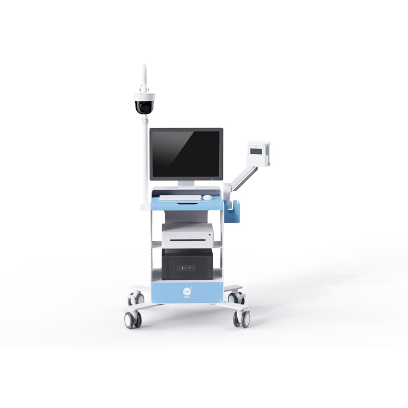
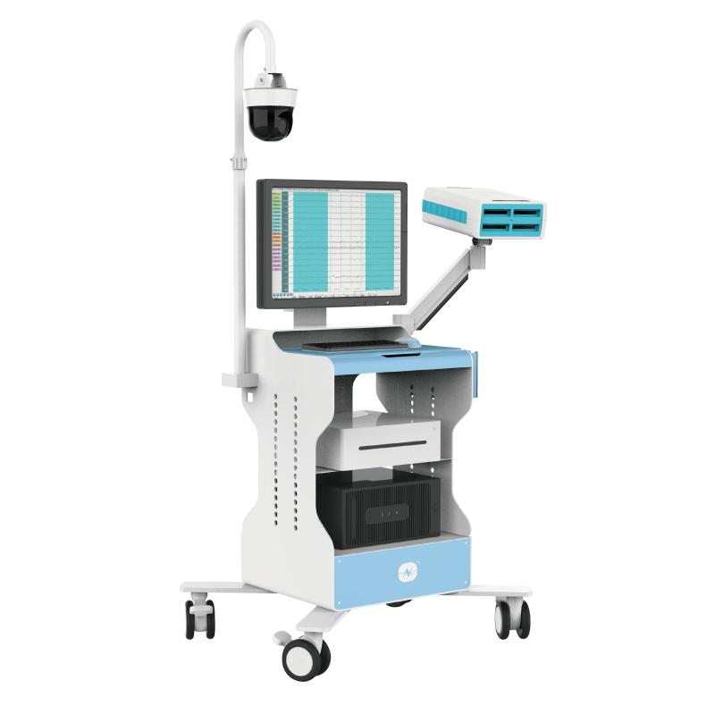
The monitoring setup, parameters, monitoring points, and intraoperative findings for each modality are described below.
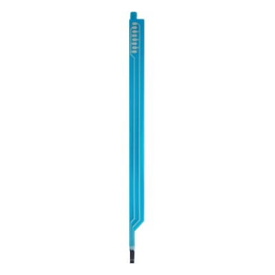
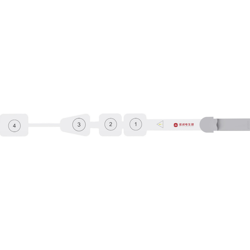
Recording electrodes: Recording electrodes were generally placed over the orbicularis oculi and orbicularis oris or mentalis muscles.
Stimulation electrodes: Stimulation was generally applied to the temporal branch, marginal mandibular branch, and supraorbital/frontal branch region of the facial nerve. The source text notes that the supraorbital/frontal branch is used less frequently and that waveform memory may be lost.
Recording parameters
| Parameter | Setting |
|---|---|
| Low-frequency filter | 30 Hz |
| High-frequency filter | 3,000 Hz |
| Sweep speed | 8 ms |
| Sensitivity | 20 microvolts |
Stimulation parameters
| Parameter | Setting |
|---|---|
| Stimulation frequency | 1 Hz |
| Stimulation intensity | 20 mA |
Before surgery, it is necessary to confirm that a typical AMR waveform can be recorded from the patient. AMR should then be monitored continuously throughout the procedure, with careful attention to changes in waveform morphology and amplitude.
In most cases, when the offending vessel has been completely removed from the site of facial nerve compression, the AMR disappears immediately or its amplitude decreases substantially, generally by more than 50%. This finding suggests that decompression has been adequate.
If the AMR does not disappear after decompression, or if it reappears after initially disappearing, the finding should be interpreted together with the Z-L response (ZLR) to determine whether an offending vessel has been overlooked or whether decompression remains incomplete. The surgeon should then re-explore the operative field and adjust the decompression as necessary.
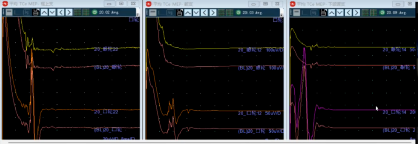
AMR waveform recorded before dural opening; the baseline was established at this stage
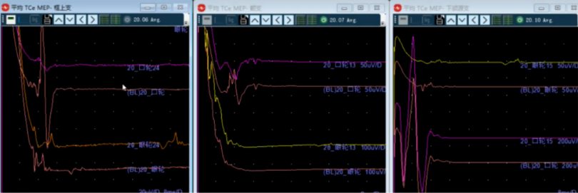
After dural opening, the waveform amplitude increased compared with the pre-dural-opening recording, and the baseline was re-established. After cerebrospinal fluid release, the supraorbital/frontal branch waveform decreased substantially
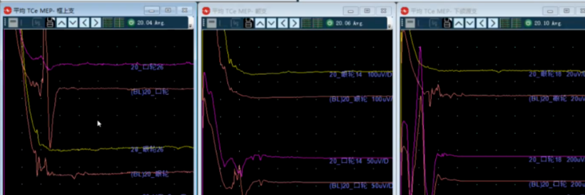
After the offending vessel was identified, the marginal mandibular branch waveform nearly disappeared
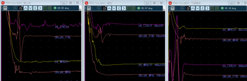
Preparation for placement of the interposition pad between the offending vessel and the facial nerve
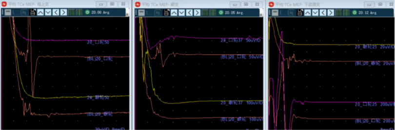
After pad placement, lateral spread responses from the supraorbital/frontal and marginal mandibular branches disappeared substantially. A residual response remained in the zygomatic branch, but its amplitude decreased markedly. The lead surgeon confirmed separation of the offending vessel and inspection of the surrounding small vessels
Recording electrodes: Scalp recording electrodes were placed according to the international 10-20 EEG system. The vertex electrode, Cz, was generally used as the positive electrode, and the preauricular electrodes, A1 and A2, were used as the negative electrodes.
Stimulation electrodes: Acoustic stimulation was delivered through sound stimulation.
Recording parameters
| Parameter | Setting |
|---|---|
| Low-frequency filter | 200 Hz |
| High-frequency filter | 2,000 Hz |
| Notch filter | Off |
| Sweep speed | 1 ms |
| Sensitivity | 0.2 microvolts |
Stimulation parameters
| Parameter | Setting |
|---|---|
| Stimulation frequency | 11.1 Hz |
| Stimulus sound polarity | Alternating phase |
| Sound mode | Short tone |
| Noise intensity mode | Absolute |
| Noise difference | 60 dB |
| Stimulation intensity | 120 dB |
BAEP recordings may contain five to seven upward-pointing peaks. Waves I, III, and V are generally the most stable and are commonly used for intraoperative monitoring. Each waveform corresponds to a specific anatomical component of the brainstem auditory pathway. BAEP therefore reflects not only conduction through the auditory pathway but also provides an indirect assessment of overall brainstem function.
During surgery, particular attention should be paid to the latency, amplitude, and morphology of waves I, III, and V. A latency prolongation of more than 10% or an amplitude reduction of more than 50% requires active investigation. Disappearance of wave V may indicate serious injury to the auditory nerve and is a highly specific warning of postoperative hearing loss or hearing impairment.
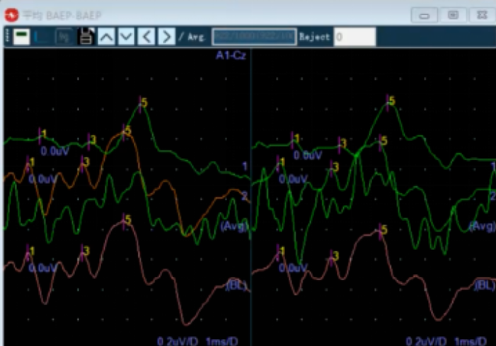
A stable BAEP waveform was recorded before dural opening and a baseline was established
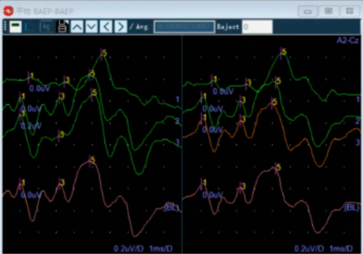
BAEP waveform recorded after release of cerebrospinal fluid
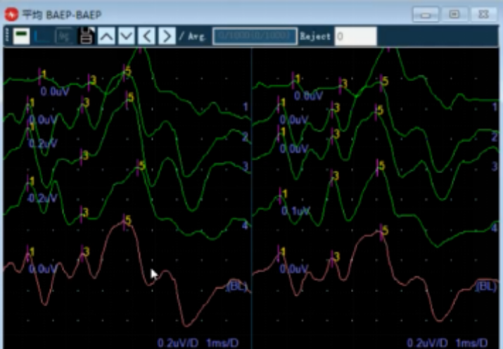
BAEP waveform recorded after identification of the offending vessel
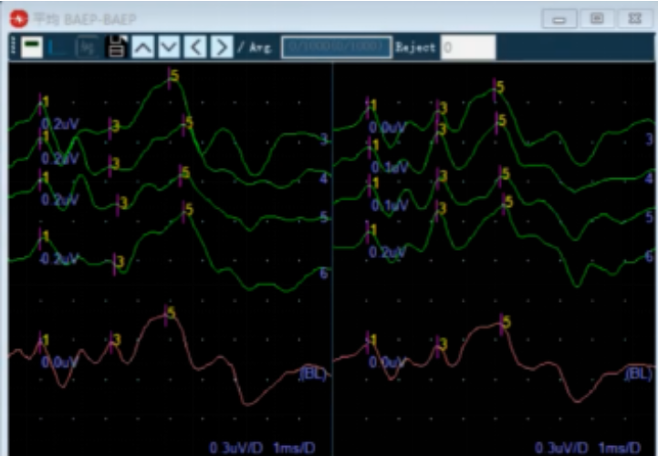
BAEP waveform recorded after placement of the interposition pad
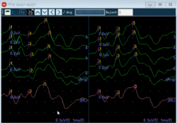
BAEP waveform monitored during wound closure
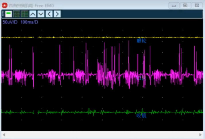
Recording electrodes: Recording electrodes were generally placed over the orbicularis oculi, orbicularis oris or mentalis, and masseter muscles.
| Parameter | Setting |
|---|---|
| Low-frequency filter | 30 Hz |
| High-frequency filter | 3,000 Hz |
| Sweep speed | 100 ms |
| Sensitivity | 50 microvolts |
EMG was mainly used to assess facial nerve motor function, help avoid injury to the motor branches of the facial nerve during surgery, and reduce the risk of postoperative facial paralysis, particularly during surgical maneuvers that could affect the facial nerve motor fibers.
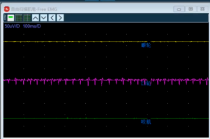
EMG response associated with mechanical traction of the facial nerve during identification of the offending vessel
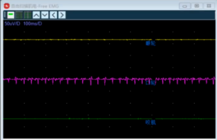
Marked EMG activity associated with facial nerve traction during separation of the nerve and vessel
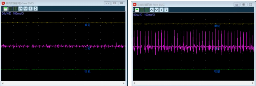
Prolonged traction was associated with sustained muscle electrical activity
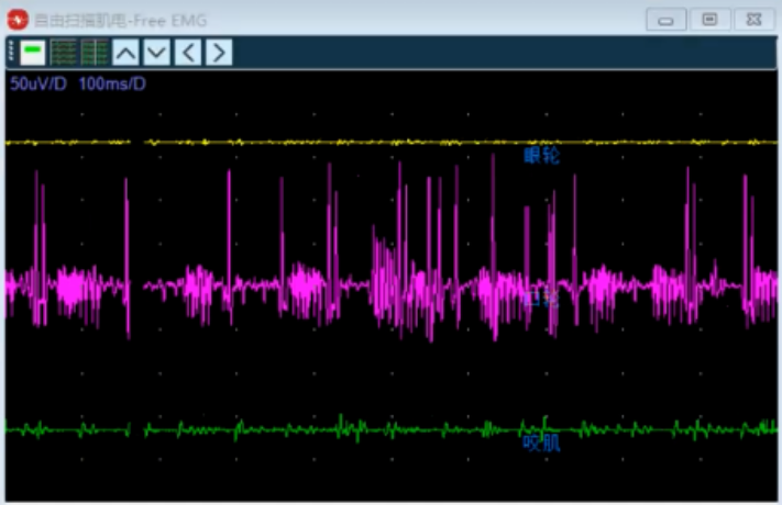
Mechanical traction during placement of the interposition pad produced an EMG response
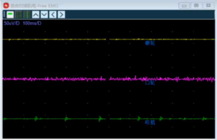
Figure caption: After placement of the interposition pad, EMG activity gradually returned to a resting state
During skin incision and craniotomy, the EMG signal showed resting electrical activity. Stable BAEP waveforms were also recorded. AMR waveforms could be elicited only with a stimulation intensity of 20 mA.
In common electrophysiological monitoring for hemifacial spasm, an AMR waveform can often be elicited at approximately 10 mA. In this patient, however, the required threshold was relatively high. Baselines were established to allow comparison throughout the operation.
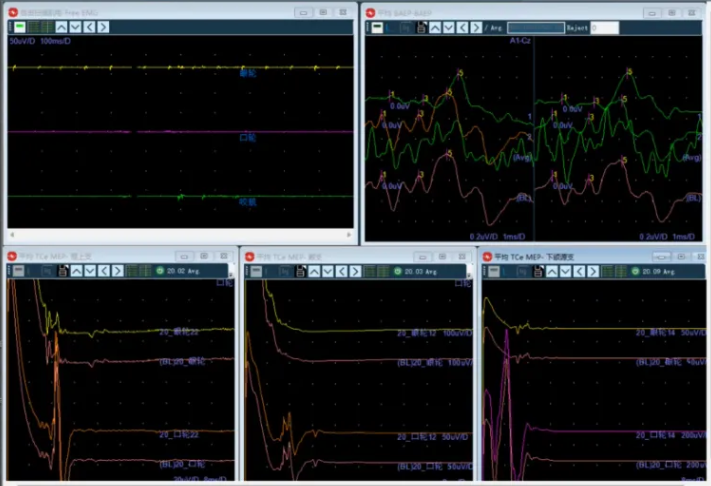
When the offending vessel was identified, the supraorbital/frontal branch waveform decreased substantially. This indicated that compression of the facial nerve by the offending vessel had been relieved to some extent.
At the same time, the facial nerve was subjected to a degree of stimulation or traction, and abnormal EMG discharges appeared. These findings indicated facial nerve stimulation. The surgeon was required to stop the relevant maneuver immediately and adjust the operative trajectory to avoid potentially irreversible neural injury.
BAEP showed no significant change during this stage.
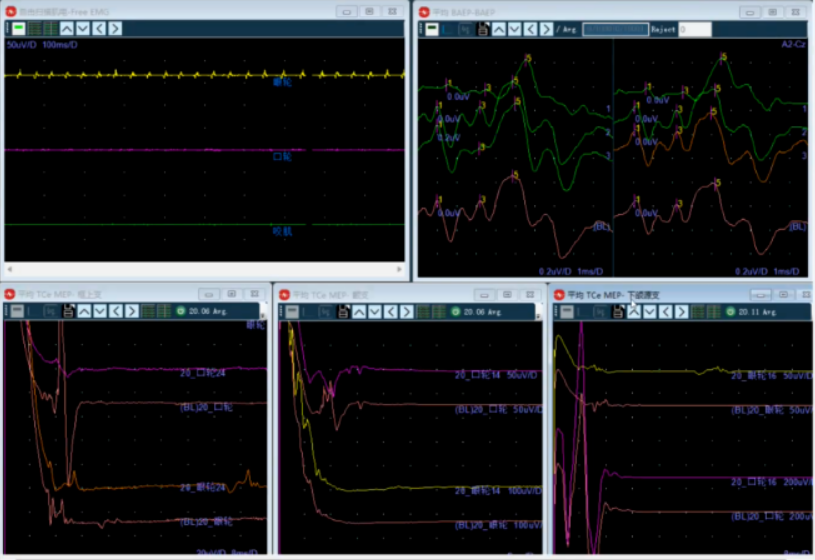
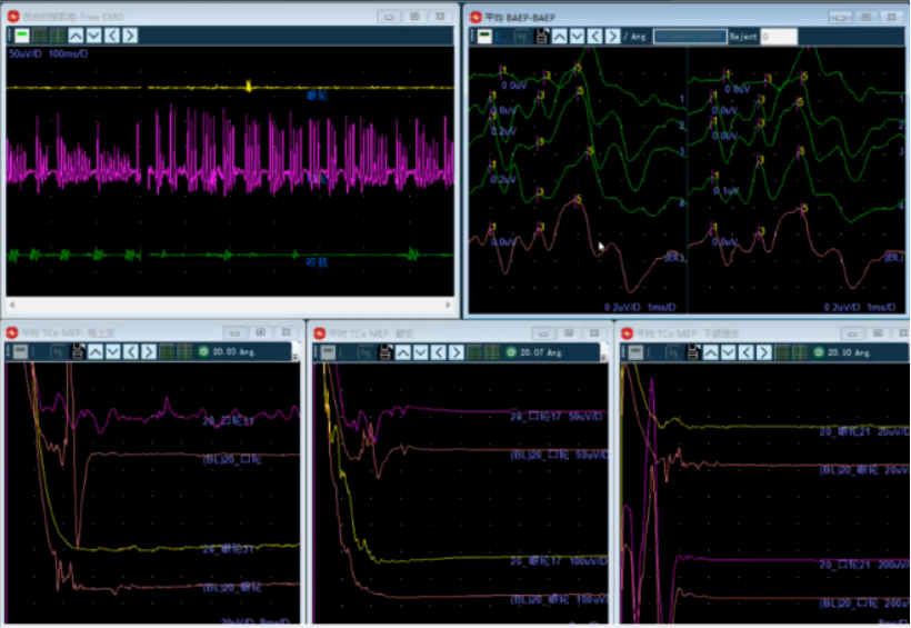
During placement of the interposition pad, the facial nerve was stimulated and abnormal EMG discharges occurred.
After the pad was placed, the lateral spread responses from the supraorbital/frontal and marginal mandibular branches disappeared. The zygomatic branch waveform decreased substantially but remained partially present. These changes indicated that pad placement had materially improved separation between the offending vessel and the facial nerve.
BAEP still showed no significant change.
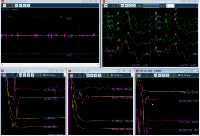
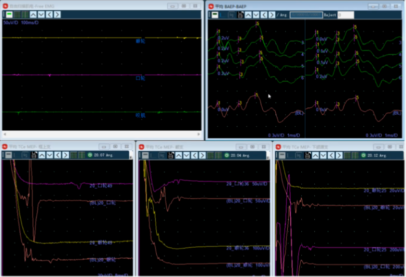
After the lead surgeon confirmed separation of the offending vessel from the facial nerve and inspection of the surrounding small vessels, the operative field was considered to have no remaining site requiring further decompression.
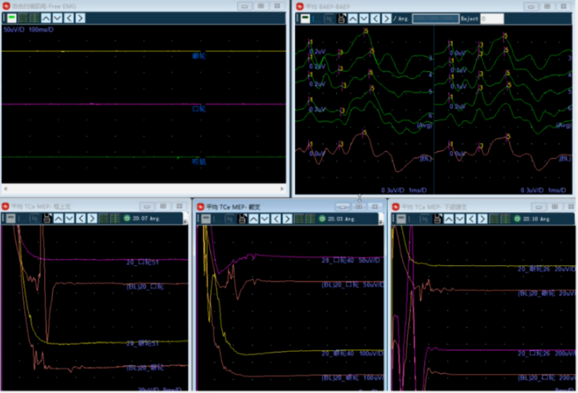
The final AMR and BAEP findings showed the following:
Intraoperative monitoring during MVD for hemifacial spasm places important demands on anesthetic management. Anesthetic agents can affect neurophysiological signals and reduce the accuracy of monitoring. Preoperative communication with the anesthesiologist is therefore essential.
Neuromuscular blocking agents should be avoided whenever possible because they may suppress EMG and AMR waveforms. During surgery, anesthetic depth should be balanced against the need for adequate sedation. Excessive anesthetic depth may suppress brainstem function and cause abnormalities in BAEP waveforms.
Changes in EMG, BAEP, and AMR across different surgical stages and maneuvers can reflect the effect of surgical manipulation on neural function. When a maneuver such as traction or electrocautery stimulates the facial nerve, abnormal EMG activity, including burst potentials, may appear. This indicates facial nerve stimulation. The surgeon should immediately stop the relevant maneuver and adjust the surgical trajectory to avoid irreversible neural injury.
In this case, BAEP waveforms showed no significant change during surgery. This finding indicated that the procedure did not cause apparent injury to the auditory conduction pathway and indirectly suggested that overall brainstem function was preserved.
At the same time, the AMR waveform decreased after cerebrospinal fluid release, identification of the offending vessel, and placement of the interposition pad. The response did not recur during closure. These findings were consistent with relief of offending-vessel compression of the facial nerve.
During identification of the offending vessel and pad placement, EMG abnormalities reflected mechanical stimulation or traction of the facial nerve. The surgical team responded by adjusting the relevant operative maneuvers. At the end of the procedure, the surgeon confirmed that the offending vessel had been separated from the facial nerve and that the surrounding small vessels had been inspected, with no further site requiring decompression.
Blood pressure and heart rate should be kept stable throughout the operation. Marked blood-pressure fluctuations may reduce perfusion of the brainstem and neural tissue and affect neurophysiological signals. Intraoperative temperature should be strictly controlled because hypothermia may slow nerve-conduction velocity and interfere with monitoring indices.
Electromagnetic interference generated by electrocautery should also be minimized to ensure correct operation of the monitoring equipment. Artifact control is an essential part of monitoring because it helps the team exclude changes unrelated to the surgical maneuver.
Hemifacial spasm is caused by microvascular compression of the facial nerve root exit zone, resulting in abnormal nerve excitability and paroxysmal, involuntary unilateral facial contractions. The condition can significantly affect quality of life and social functioning.
Microvascular decompression is a definitive surgical treatment for hemifacial spasm. Its core objectives are to identify and relieve compression of the facial nerve by the offending vessel while protecting the facial nerve, auditory nerve, and brainstem to the greatest possible extent.
Intraoperative neurophysiological monitoring provides an objective and real-time assessment of neural function under anesthesia. It functions as an additional layer of neurological protection during surgery. AMR/LSR can assist in evaluating the adequacy of decompression and may provide early warning of persistent or recurrent facial nerve compression. BAEP is primarily used to monitor auditory pathway and brainstem function, while EMG helps detect mechanical traction or thermal stimulation of the facial nerve and its motor branches.
In this case, multimodal monitoring supported completion of decompression while helping avoid apparent injury to facial or auditory neural function. The procedure was associated with disappearance or marked reduction of the relevant AMR/LSR responses, stable BAEP waves III and V, and gradual return of EMG activity to resting levels in the absence of stimulation.
The case illustrates the value of combining AMR/LSR, BAEP, and EMG during MVD for hemifacial spasm. Continuous waveform assessment, prompt communication, careful handling of the facial nerve, and systematic control of anesthetic, hemodynamic, temperature, and artifact-related factors all contribute to safer intraoperative decision-making.

Copyright © NCC Medical Co., Ltd. - Privacy policy
 EN