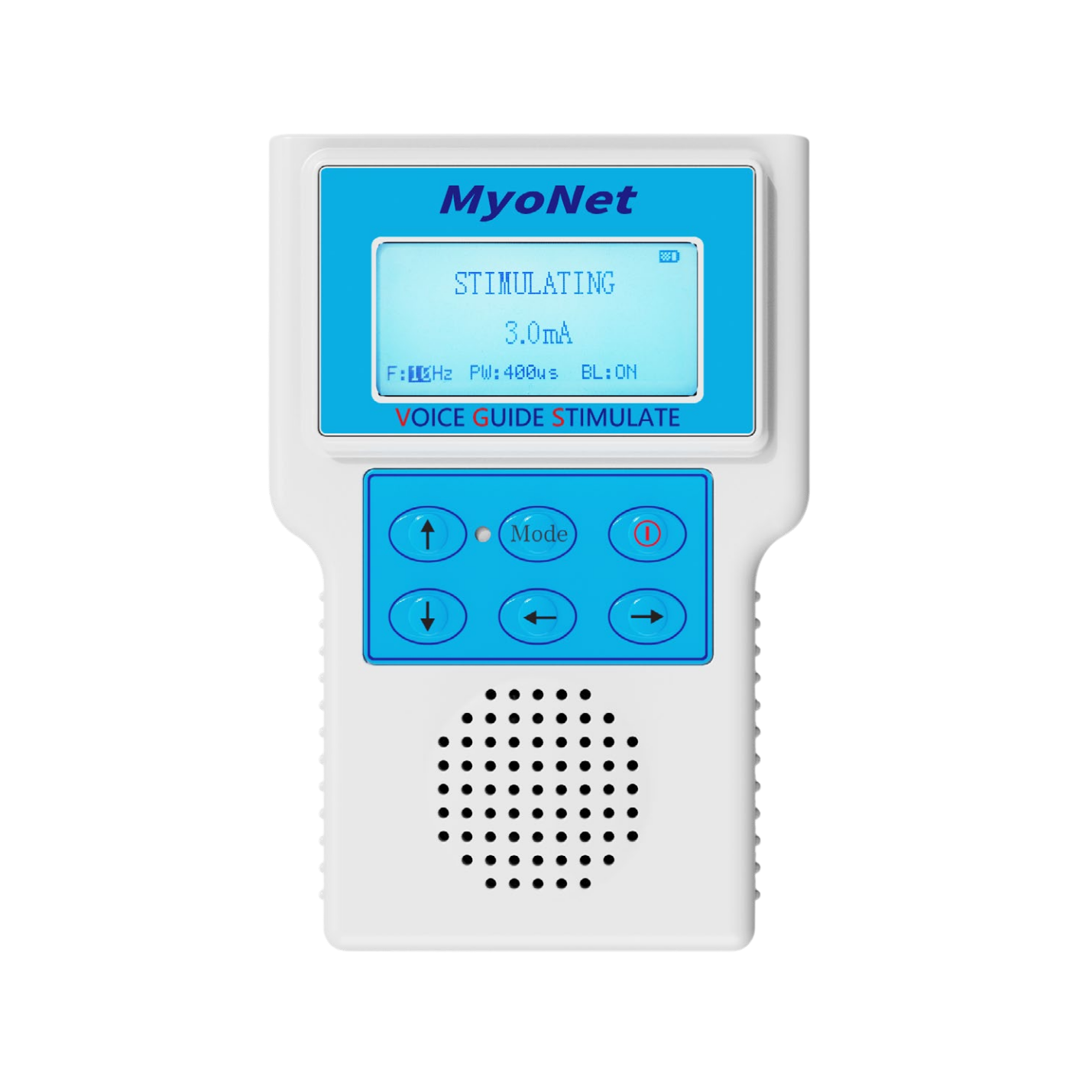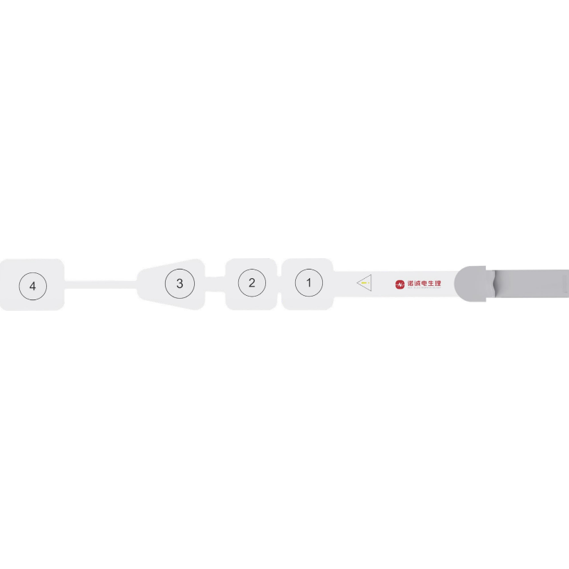
This case report describes multimodal intraoperative neurophysiological monitoring during UBE surgery for L4-L5 lumbar disc herniation, including EMG, TCeMEP, and SEP monitoring during decompression and thermal annuloplasty.
A 48-year-old man underwent unilateral biportal endoscopy (UBE) surgery for an L4-L5 lumbar disc herniation. The procedure included removal of the herniated nucleus pulposus, lumbar nerve-root decompression, and thermal annuloplasty of the annulus fibrosus.
Before surgery, the patient’s main symptoms were lower-extremity numbness and pain. Lower-extremity muscle strength was normal on preoperative examination.
The objectives of surgery were to remove the herniated nucleus pulposus, relieve compression of the affected nerve root, and alleviate the patient’s lower-extremity numbness and pain. Thermal treatment of the annular defect was also performed with the aim of reducing the risk of postoperative recurrence.
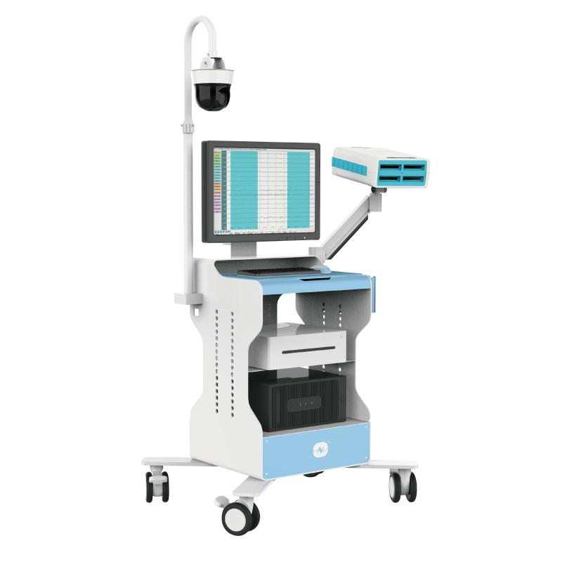
The neurophysiological monitoring modalities commonly used during UBE surgery include spontaneous electromyography (EMG), somatosensory evoked potentials (SEP), and transcranial electrical motor evoked potentials (TCeMEP).
Among these modalities, EMG is one of the most specific and clinically important monitoring indicators during UBE procedures because it can provide real-time warning of mechanical stimulation or thermal injury to the nerve root. SEP and TCeMEP provide complementary information regarding sensory and motor pathway function.
The monitoring findings and parameters for each modality are described below.
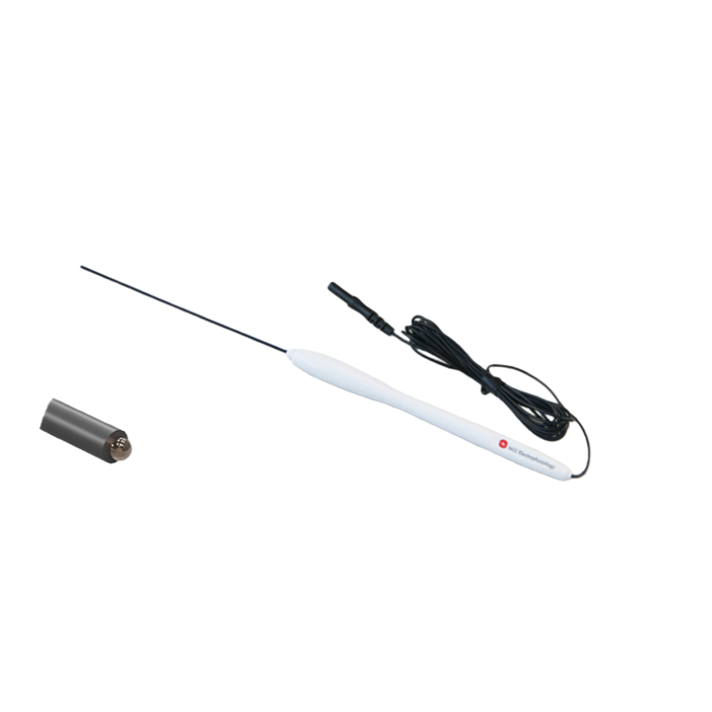
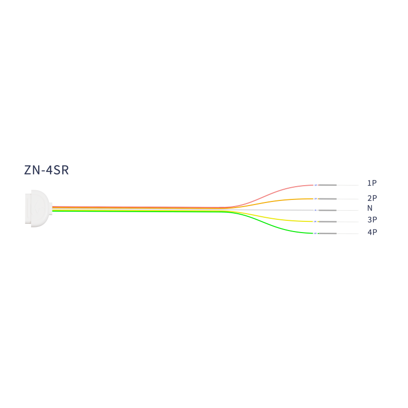
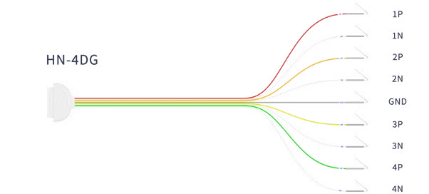
Recording electrodes: Recording electrodes were generally placed over the quadriceps, tibialis anterior, and abductor hallucis muscles.
Recording parameters
| Parameter | Setting |
|---|---|
| Low-frequency filter | 30 Hz |
| High-frequency filter | 3,000 Hz |
| Sweep speed | 100 ms |
| Sensitivity | 200 microvolts |
Reported alert criteria: A reduction in amplitude of at least 50% from baseline or a latency prolongation of at least 10%, when sustained for at least 3 minutes, required urgent management according to the source case description.
EMG was used to provide real-time warning of mechanical stimulation and thermal injury to the nerve root, to help protect the patient’s normal preoperative motor function, and to reduce the risk of postoperative neurological complications.
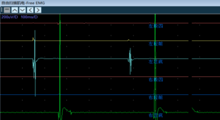
Free-run EMG was continuously observed during surgical manipulation. Mechanical stimulation of the nerve root could produce abnormal spontaneous activity, including trains of discharges or burst activity. Such activity required immediate notification of the surgical team and reassessment of the relevant maneuver.
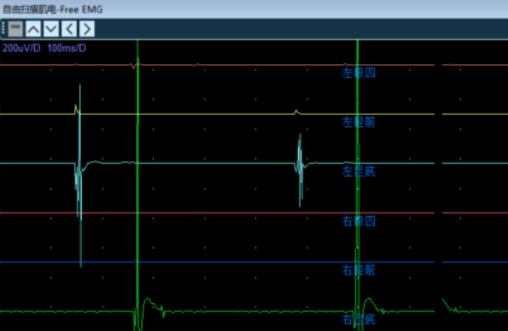
Free-run EMG recorded during mechanical stimulation
Stimulation electrodes: In accordance with the international 10-20 EEG system, stimulation electrodes were placed at C3 and C4 on the scalp.
Recording electrodes: Recording electrodes were generally placed bilaterally over the quadriceps, tibialis anterior, and abductor hallucis muscles.
| Parameter | Setting |
|---|---|
| Low-frequency filter | 30 Hz |
| High-frequency filter | 3,000 Hz |
| Notch filter | Off |
| Sweep speed | 10 ms |
| Sensitivity | 50 microvolts |
| Parameter | Setting |
|---|---|
| Stimulation frequency | 0.1 Hz |
| Pulse width | 75 microseconds |
| Number of pulses per train | 7 |
| Train frequency | 500 Hz |
| Stimulation intensity | 200-400 V |
TCeMEP was used to provide real-time warning of possible mechanical stimulation or thermal injury affecting the nerve root and to help preserve the patient’s normal preoperative motor function, thereby reducing the risk of postoperative neurological complications.
A stable TCeMEP baseline should be obtained before decompression. Continuous intraoperative monitoring should then be maintained, and subsequent waveform changes should be compared with the preoperative baseline.
According to the source case, an amplitude reduction of more than 50% from baseline or a latency prolongation of more than 10% was used as an alarm criterion. Neuromuscular blocking agents and excessive anesthetic depth should be avoided because they may suppress the TCeMEP waveform. When an abnormal change is identified, the monitoring team should issue a timely alert so that the surgical team can investigate and respond.
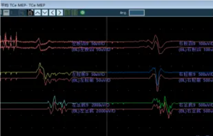
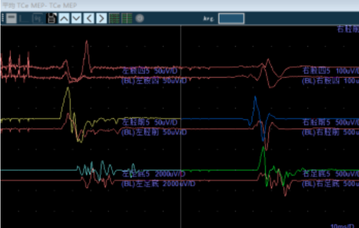
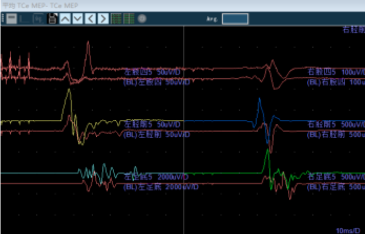
A stable TCeMEP waveform was obtained before decompression and a baseline was established
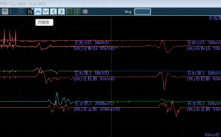
TCeMEP waveform recorded after additional neuromuscular blockade
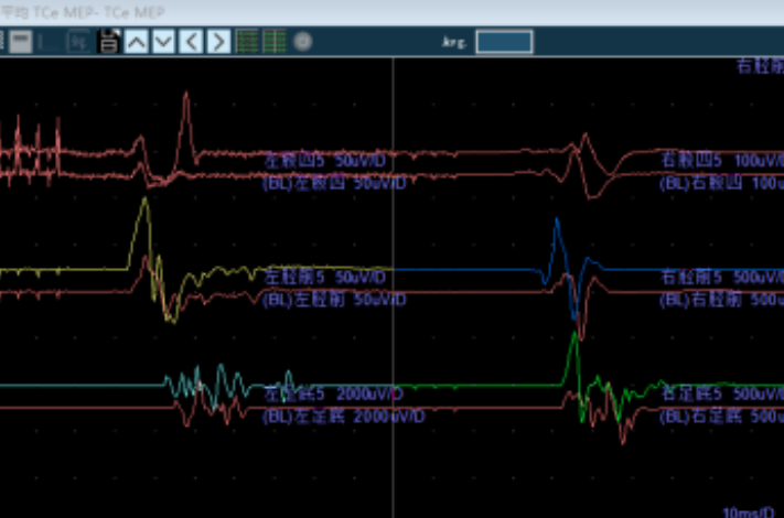
TCeMEP waveform recorded after removal of the herniated nucleus pulposus
Recording electrodes: In accordance with the international 10-20 EEG system, the recording electrode was placed at Cz', approximately 2 cm posterior to Cz. The reference electrode was placed at Fz.
Stimulation electrodes: For lower-extremity stimulation, the stimulating electrodes were placed at the medial ankle over the posterior tibial nerve.
| Parameter | Setting |
|---|---|
| Low-frequency filter | 30 Hz |
| High-frequency filter | 500 Hz |
| Sweep speed | 10 ms |
| Sensitivity | 2 microvolts |
| Parameter | Setting |
|---|---|
| Stimulation frequency | 2.33 Hz |
| Pulse width | 200 microseconds |
| Stimulation intensity | 25 mA |
SEP was used to assess the integrity of the sensory conduction pathways and to help prevent postoperative sensory dysfunction.
A stable SEP baseline should be obtained before surgery and monitored continuously throughout the procedure. According to the source case, an amplitude reduction of more than 50% from baseline or a latency prolongation of more than 10% was used as an alarm criterion.
Excessive anesthetic depth and hypotension should be avoided because they may interfere with SEP signals. If an abnormal change is observed, the monitoring team should issue a timely alert and the possible causes should be investigated.
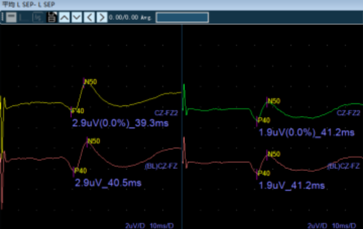
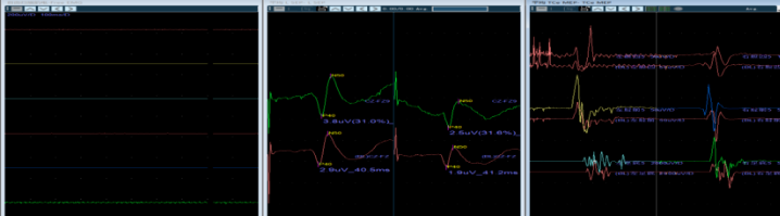
A stable SEP waveform was recorded before surgery and a baseline was established
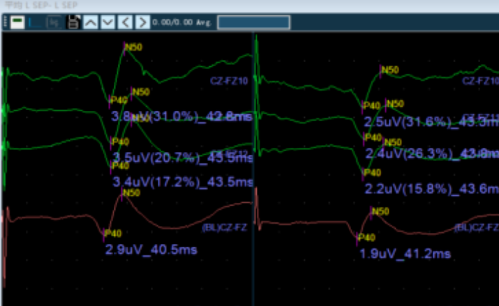
Intraoperative SEP waveform recording
At the beginning of surgery, while the UBE working channels were being established, the EMG signal showed resting electrical activity. Stable SEP and TCeMEP waveforms were also recorded. These signals were used to establish the baseline for real-time comparison during the remainder of the operation.
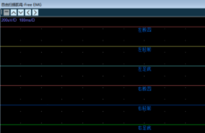
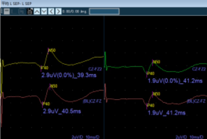
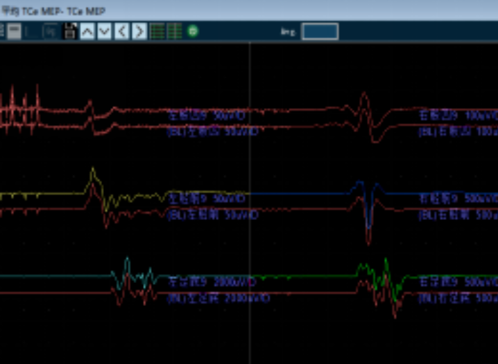
During precise decompression and treatment of the annulus fibrosus, free-run EMG was monitored continuously. The occurrence of trains of discharges or burst activity would have required an immediate alert because such activity could indicate nerve-root stimulation.
After decompression was completed, the TCeMEP amplitude was clearly higher than the preoperative baseline.
At the end of the procedure, the lead surgeon confirmed that the affected nerve root had been completely released and that there was no active bleeding.
SEP and TCeMEP were then assessed again. The final monitoring results showed that the amplitudes and latencies remained within the normal range. In the absence of stimulation, EMG continued to show resting electrical activity.
Anesthesia management is particularly important during intraoperative monitoring for lumbar UBE surgery. Anesthetic agents may affect neurophysiological signals and compromise the accuracy of monitoring. Preoperative communication with the anesthesiologist is therefore necessary.
Neuromuscular blocking agents and excessive anesthetic depth should be avoided because they may suppress TCeMEP waveforms. Anesthetic management should be coordinated with the monitoring requirements so that reliable motor and sensory signals can be obtained and maintained.
During surgery, free-run EMG, TCeMEP, and SEP should be monitored continuously. When a surgical maneuver, such as nerve-root traction or electrocautery, stimulates the nerve root, abnormal EMG activity, including burst potentials, may occur. Such a change suggests that the nerve root may be undergoing mechanical or thermal stimulation. The surgeon should immediately review and adjust the surgical trajectory or maneuver to avoid neural injury.
In this case, the TCeMEP and SEP amplitudes and latencies did not show material changes compared with the preoperative baseline. No alarm-level amplitude reduction of more than 50% or latency prolongation of more than 10% was observed. After decompression, TCeMEP amplitude was higher than the preoperative baseline.
Blood pressure, heart rate, and body temperature should be maintained within stable ranges throughout the procedure. Electromagnetic interference should also be minimized to ensure reliable operation of the monitoring equipment and to reduce artifacts that could be mistaken for physiological changes.
The monitoring results were interpreted together with the surgical findings. After the surgeon confirmed complete nerve-root release and the absence of active bleeding, the final SEP and TCeMEP recordings remained within the normal range, while EMG remained at resting activity without stimulation.
Lumbar disc herniation occurs when nucleus pulposus tissue protrudes into the spinal canal and compresses a nerve root, causing lower-extremity numbness and pain. In severe cases, these symptoms can affect walking ability and quality of life.
UBE-assisted removal of the herniated nucleus pulposus combined with nerve-root decompression and thermal annuloplasty is a minimally invasive surgical approach for lumbar disc herniation. It is intended to directly remove the herniated tissue, relieve nerve-root compression, and treat the annular defect.
Intraoperative neurophysiological monitoring provides an objective and real-time assessment of neural function under anesthesia. It functions as an additional layer of neurological protection during surgery. In particular, free-run EMG can provide real-time warning of mechanical or thermal stimulation of the nerve root, while TCeMEP and SEP provide complementary information about motor and sensory pathway function.
In this case, multimodal monitoring helped support the safe completion of precise nucleus pulposus removal and adequate nerve-root decompression without evidence of intraoperative neurological deterioration. The monitoring record showed stable SEP and TCeMEP responses relative to the preoperative baseline, an increase in TCeMEP amplitude after decompression, and resting EMG activity in the absence of stimulation.
These findings supported the overall surgical objective of relieving nerve-root compression while protecting neurological function and providing a favorable basis for postoperative recovery and quality-of-life improvement.

Copyright © NCC Medical Co., Ltd. - Privacy policy
 EN