This case report describes multimodal intraoperative neurophysiological monitoring during L4-L5 fusion and decompression for lumbar disc herniation with radiculopathy, focusing on SSEP, TcMEP, free-run EMG, and graded responses to intraoperative neurological alerts.
A 37-year-old woman was preoperatively diagnosed with lumbar disc herniation accompanied by radiculopathy.
The surgical objective was to improve the patient's body alignment and restore quality of life through L4-L5 fusion and decompression.
L4-L5 lumbar fusion and decompression—whether performed as posterior lumbar interbody fusion (PLIF), transforaminal lumbar interbody fusion (TLIF), or open decompression with pedicle-screw fixation—primarily involves the L4 and L5 nerve roots, the cauda equina, and the lumbosacral plexus.
Potential intraoperative risks include:
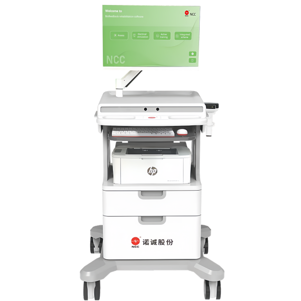
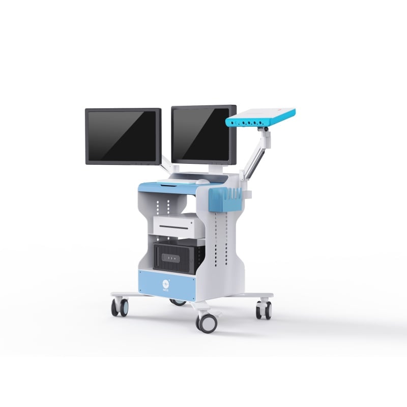
The standard monitoring protocol consists of lower-extremity somatosensory evoked potentials (SSEP), lumbosacral motor evoked potentials (MEP), and free-run electromyography (f-EMG). Anal sphincter EMG may be added when clinically necessary.
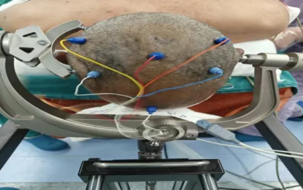
SSEP was used to monitor the posterior columns of the spinal cord, lumbosacral sensory conduction, and the sensory fibers of the cauda equina.
An amplitude reduction of at least 50% or a latency prolongation of at least 10%, sustained for more than 3 minutes and occurring after technical factors have been excluded, was treated as an alert.
SSEP changes may provide an early warning of cauda equina compression, lumbosacral ischemia, or excessive traction.
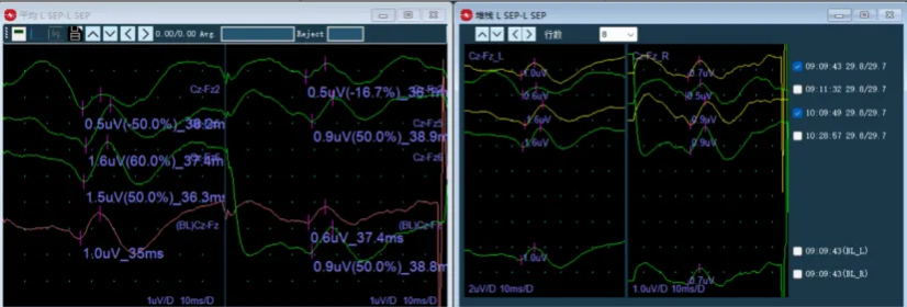
Lower-extremity SSEP waveforms were recorded throughout the operation. No abnormality was reported during the procedure, which was completed quickly by the surgeon.
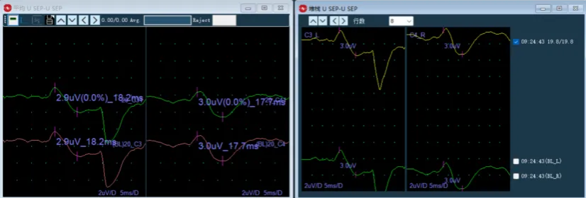
The source text states that USEP was recorded as a reference for LSEP.
| Parameter | Setting |
|---|---|
| Stimulation frequency | 2.33 Hz |
| Stimulation intensity | 25 mA, constant current |
| Pulse type | Single pulse |
| Pulse width | 200 microseconds |
TcMEP was used to monitor the corticospinal tracts, lumbosacral motor nerve roots, and motor fibers of the cauda equina.
A reduction in amplitude of at least 80% or complete disappearance of the response was treated as an alert.
TcMEP was used as an early warning of motor injury and is highly sensitive to changes occurring during decompression, pedicle-screw placement, and interbody-cage implantation.
After fusion and decompression were completed, motor-evoked-potential waveforms were recorded. The source case reports that the operation proceeded smoothly without abnormal intraoperative findings.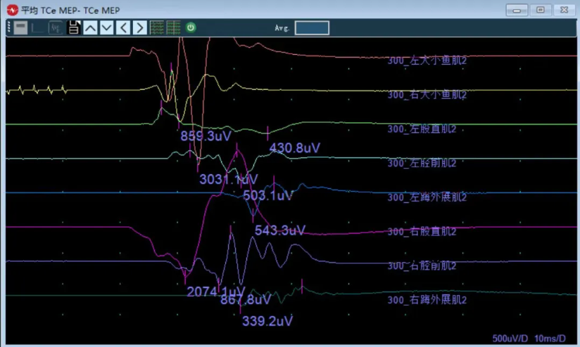
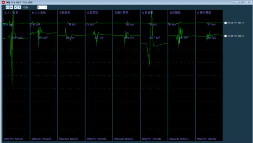
| Parameter | Setting |
|---|---|
| Pulse type | Single pulse train |
| Number of pulses per train | 5 |
| Pulse width | 75 microseconds |
| Train frequency | 1,000 Hz |
| Stimulation intensity | 220 V |
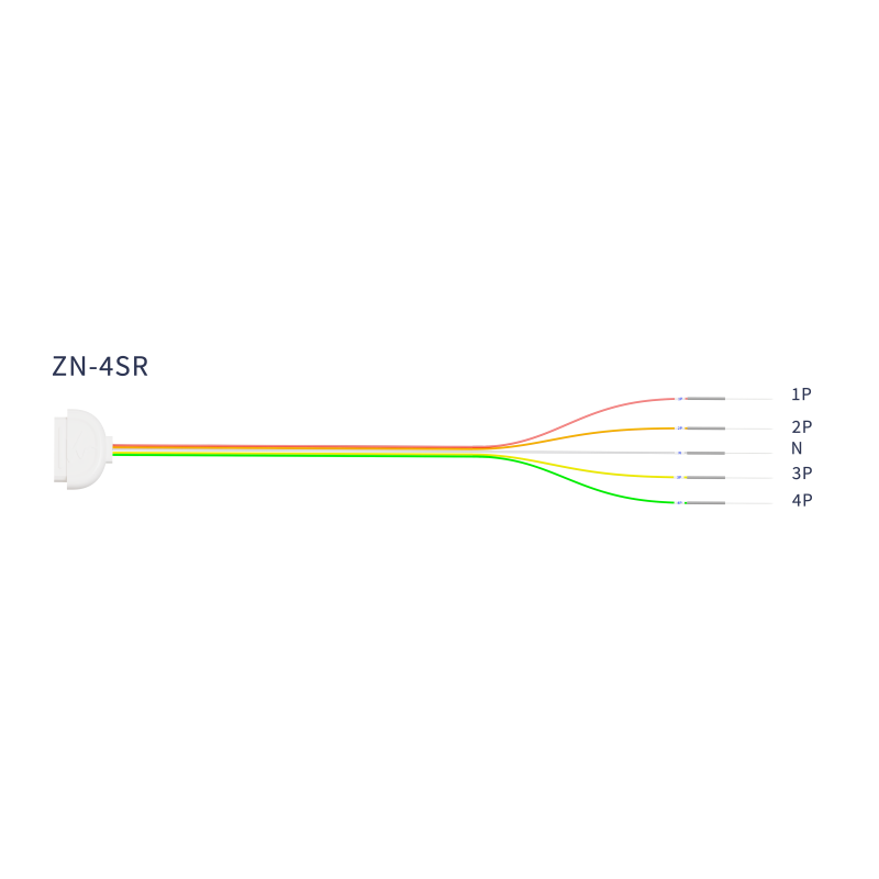
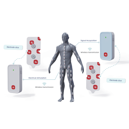
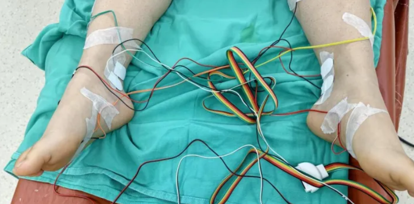
f-EMG was recorded from the bilateral tibialis anterior, rectus femoris, abductor hallucis, thenar, and hypothenar muscles.
The evoked-potential system, electrodes, and leads should be checked before surgery. Potential interference from electrocautery and powered drills should also be assessed.
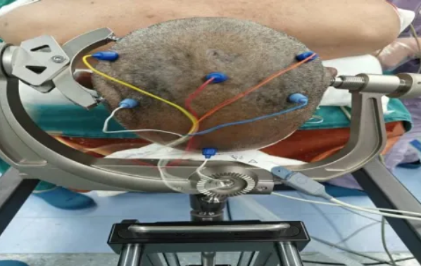
After positioning has been completed and anesthesia is stable, obtain bilateral SSEP, MEP, and resting EMG recordings. Save the baseline and repeat the acquisition twice to confirm stability.
Risk: Retractor-related compression or excessive traction on the L4-L5 nerve roots.
Monitoring response: Burst potentials should prompt the monitoring team to remind the surgeon to relax the retractors intermittently.
Risks:
Monitoring focus: Observe bilateral SSEP and MEP waveforms in real time.
Critical maneuvers requiring particular attention:
A unilateral MEP decrease may indicate a nerve-root problem and requires prompt assessment.
Triggered EMG (t-EMG) should be monitored throughout the sequence of pedicle access, probing, tapping, and screw insertion.
After each screw has been placed, measure and document its stimulation threshold. A threshold below 10 mA requires immediate feedback to the surgical team and reassessment or adjustment of the screw trajectory.
Risks:
Monitoring focus: TcMEP and EMG.
Post-implantation reassessment: Repeat monitoring after cage placement. The position should be considered acceptable only when there is no obvious attenuation of the monitoring waveforms.
Risks: Excessive compression causing nerve-root traction or excessive tension on the cauda equina.
Monitoring focus: Continue MEP and SSEP monitoring and avoid excessive reduction or correction.
Repeat the complete SSEP, MEP, and EMG assessment and compare the findings with the baseline.
The wound should be closed only after bilateral waveforms remain stable without significant attenuation.
At the end of the procedure:
Finding: Intermittent, scattered EMG discharges.
Response: Provide a verbal alert, continue close observation, and proceed with surgery while monitoring the trend.
Finding: Clustered or burst EMG activity, or mild SSEP latency prolongation.
Response: Pause the local surgical maneuver, adjust the retractor or instrument, and repeat the monitoring assessment.
Finding: SSEP amplitude reduction of at least 50%, or MEP amplitude reduction of at least 80% or complete loss of the response.
Response:
Abdominal compression in the prone position may impair venous return and contribute to venous congestion of the cauda equina. The baseline should therefore be rechecked after positioning.
Use the drill intermittently and apply water cooling as appropriate to reduce heat- and vibration-related EMG artifacts.
Deep anesthesia during surgery should be strictly avoided because it may cause a false reduction in MEP or SSEP amplitudes.
When an abnormality is identified, the surgeon and anesthesiologist should be informed immediately, and intervention should begin within 3 minutes.
The core of IONM during L4-L5 lumbar fusion and decompression is continuous monitoring with lower-extremity SSEP, lumbosacral MEP, and free-run EMG, with particular attention to protection of the L4 and L5 nerve roots and the cauda equina.
When combined with TIVA, rechecking the baseline after prone positioning, and rapid graded intervention for abnormalities, this monitoring strategy is intended to reduce the risk of nerve-root and cauda equina injury and to help avoid postoperative lower-extremity weakness, numbness, and bowel or bladder dysfunction.

Copyright © NCC Medical Co., Ltd. - Privacy policy
 EN