A case report of multimodal intraoperative neurophysiological monitoring during congenital scoliosis correction, highlighting TcMEP loss during correction, prompt surgical adjustment, and waveform recovery.
A 10-year-old boy had a 9-year history of scoliosis with progressive worsening of the deformity. He had a pronounced left-sided cervical curvature with straightening of the normal cervical lordosis, together with a mild leftward curvature of the upper thoracic spine.
The patient underwent scoliosis correction with osteotomy. The goals of surgery were to protect spinal cord and neural function to the greatest possible extent; arrest further progression of the deformity by stabilizing the hemivertebra and other abnormal structures; correct trunk imbalance and improve appearance; and increase thoracic volume to help protect and improve cardiopulmonary function.
The neurophysiological monitoring modalities commonly used during scoliosis correction include motor evoked potentials (MEP), somatosensory evoked potentials (SEP), and electromyography (EMG). These three modalities have distinct and complementary roles:
MEP is highly sensitive to changes in motor pathway function, including amplitude reduction, and may identify isolated motor injury that could be missed when relying on SEP alone. Together, multimodal monitoring is intended to reduce the risk of intraoperative neurological injury and postoperative paralysis.
Recording electrodes: Based on the C3-C5 operative level, recording electrodes were placed over the deltoid, trapezius, thenar muscles, hypothenar muscles, and abductor hallucis muscles.
Stimulation electrodes: Transcranial stimulation electrodes were placed on the scalp over the motor cortex, commonly at C1/C2 or C3/C4 positions.
Recording parameters
| Parameter | Setting |
|---|---|
| Low-frequency filter | 30 Hz |
| High-frequency filter | 3,000 Hz |
| Sweep speed | 10 ms |
| Sensitivity | 50-500 microvolts |
Stimulation parameters
| Parameter | Setting |
|---|---|
| Stimulation frequency | 1 Hz |
| Stimulation intensity | 200-330 mA |
At the start of surgery, after the patient has been positioned and secured, stable and reproducible MEP waveforms should be obtained immediately as the baseline. All subsequent waveform changes should be compared with this patient-specific baseline rather than with absolute normative values.
MEP is highly sensitive to anesthetic depth and neuromuscular blocking agents. Total intravenous anesthesia (TIVA), such as propofol plus remifentanil, should be used. Neuromuscular blocking agents should be avoided after induction; if used, they should be limited to induction only, to maintain stable MEP signals.
Amplitude reduction: A reduction in MEP amplitude of more than 50%-80% from baseline may be considered a major warning. Different centers may use different criteria; in this case source, a reduction of 75% was described as clinically significant.
Latency prolongation: A marked prolongation in latency, such as more than 10%, may suggest demyelination or temperature-related change. It is less sensitive than amplitude change and should be interpreted as an adjunctive indicator.
Complete signal loss: Complete disappearance of MEP signals in any monitored channel, after exclusion of technical or anesthetic causes, is a serious alert. The relevant surgical maneuver should be stopped immediately and rescue measures initiated.
Pedicle screw placement: Triggered EMG should be used to assess whether a screw is approaching a nerve root and to help avoid direct neural injury.
Deformity correction: Distraction, compression, derotation, and other correction maneuvers may stretch the spinal cord. MEP trends should be monitored continuously during these steps.
Osteotomy and closure maneuvers: After spinal osteotomy, MEP amplitude may transiently decrease during closure or compression. If a decrease persists without recovery, the surgeon should reassess and adjust the procedure.
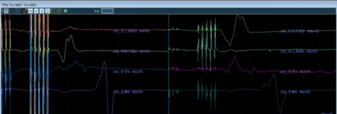
MEP waveform recorded before surgery as the baseline. No additional neuromuscular blocking agent was administered after induction.
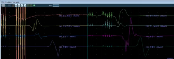
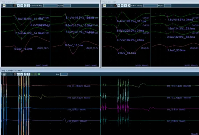
MEP monitoring during pedicle screw placement.
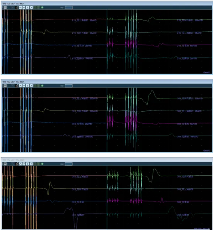
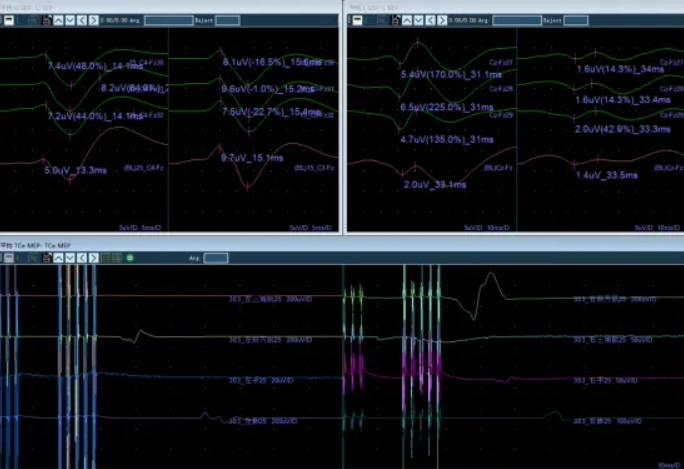
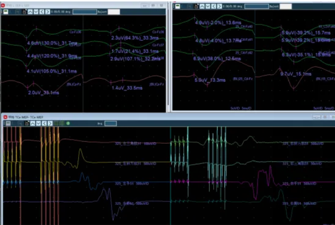
MEP showed an evident change during compressive scoliosis correction. Following notification and adjustment of the correction maneuver, the waveform gradually recovered.
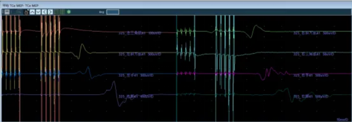
Motor responses were largely recovered during osteotomy.
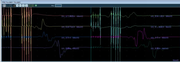
At closure, final MEP amplitudes had recovered to the preoperative level.
Recording electrodes: Electrodes were placed according to the international 10-20 system.
Stimulation electrodes:
Recording parameters
| Parameter | Upper Extremities | Lower Extremities |
|---|---|---|
| Sweep speed | 5 ms | 10 ms |
| Sensitivity | 5 microvolts | 5 microvolts |
Stimulation parameters
| Parameter | Setting |
|---|---|
| Stimulation frequency | 2.33 Hz |
| Stimulation pulse | Single-pulse train |
| Pulse polarity | Positive |
| Stimulation intensity, upper extremity | 20 mA |
| Stimulation intensity, lower extremity | 25 mA |
The SEP trace may show a series of upward peaks. The upper-extremity N20 response, recorded over the parietal cortex, and the lower-extremity P40 response, recorded over the postcentral gyrus, are generally the most stable signals used for intraoperative monitoring.
The N20 response reflects function of the thalamocortical radiation and primary somatosensory cortex. The P40 response reflects the conduction integrity of the dorsal columns and medial lemniscus. Intraoperative assessment should focus on the latency, amplitude, and morphology of N20 and P40.
A latency prolongation of more than 10% or an amplitude reduction of more than 50% from baseline requires active investigation. Complete loss of N20 or P40, after technical and anesthetic causes have been excluded, may indicate serious compromise of the dorsal columns or thalamocortical sensory pathways and is a highly specific warning of postoperative sensory dysfunction.
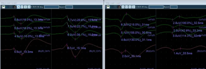
Stable BAEP waveforms were recorded and baseline measurements were established before dural opening.
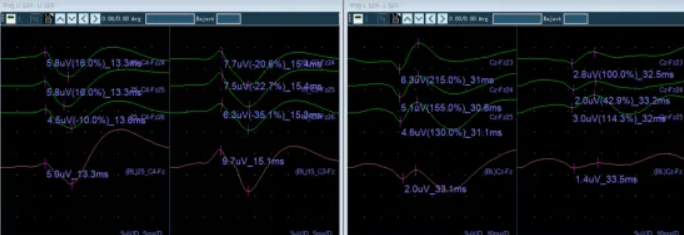
SEP monitoring during pedicle screw placement.
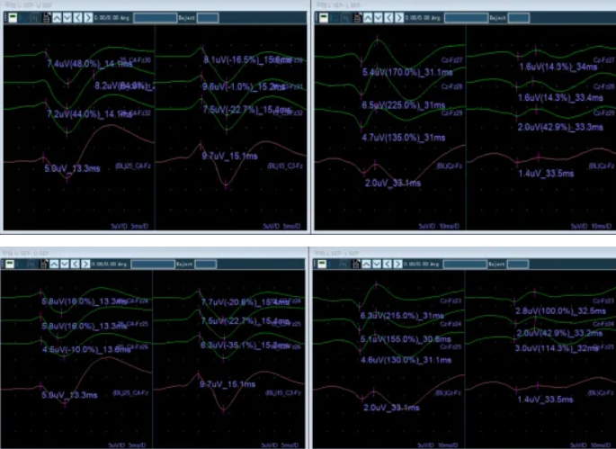
SEP monitoring during corrective decompression.
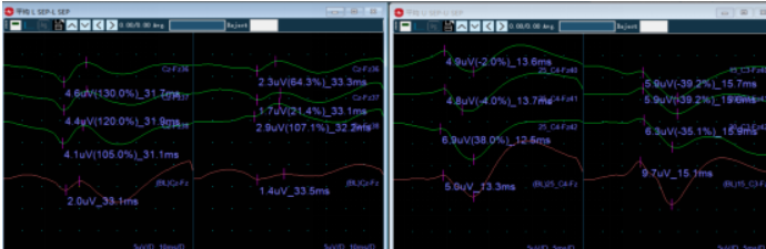
SEP monitoring during closure.
SEP showed no significant change throughout the procedure.
Recording electrodes: Electrodes were generally placed over the deltoid, trapezius, thenar muscles, hypothenar muscles, and abductor hallucis muscles.
| Parameter | Setting |
|---|---|
| Low-frequency filter | 30 Hz |
| High-frequency filter | 3,000 Hz |
| Sweep speed | 200 ms |
| Sensitivity | 50 microvolts |
EMG was used to evaluate intraoperative spinal cord and motor nerve-root function and to help avoid injury to motor neural structures during surgery, thereby reducing the risk of postoperative paralysis. Particular attention was required during pedicle screw placement, corrective distraction, and osteotomy closure, as these stages may affect spinal motor nerve roots.
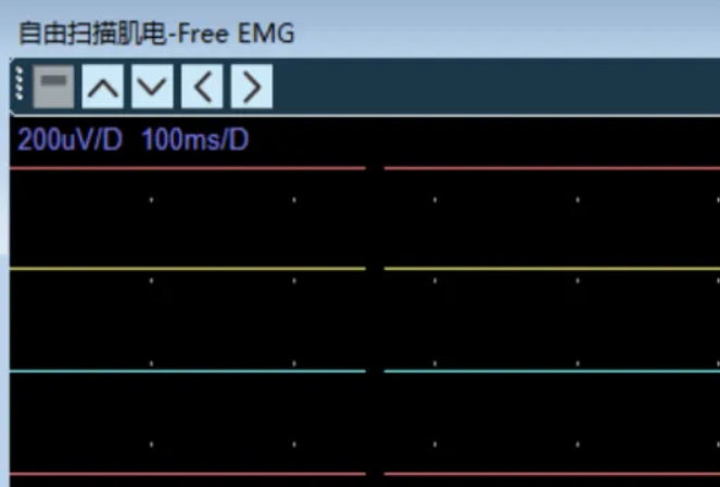
During pedicle screw placement, corrective distraction, and osteotomy closure, no clear EMG response consistent with significant traction was identified.
During skin incision, paraspinal muscle dissection, and pedicle exposure, free-run EMG showed resting electrical activity without spontaneous abnormal discharges. Stable SEP waveforms were recorded, including upper-extremity N20 and lower-extremity P40 responses with normal latency and amplitude. Clear and reproducible MEP compound muscle action potentials were elicited from the monitored target muscles of all extremities.
Baselines for SEP, MEP, and Free-EMG were established to enable real-time intraoperative comparison and warning.
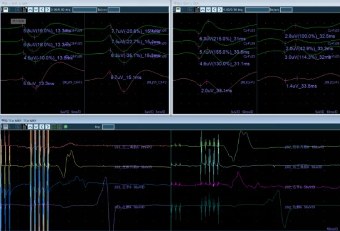
During pedicle screw placement, SEP and MEP showed no significant change. These findings suggested that screw placement did not enter the spinal canal or compress or traction the spinal cord, and that dorsal column function monitored by SEP and corticospinal tract function monitored by MEP remained intact. There was no apparent spinal cord ischemia during screw placement, with no evidence that radiculomedullary or segmental blood supply had been injured sufficiently to cause spinal cord hypoperfusion.
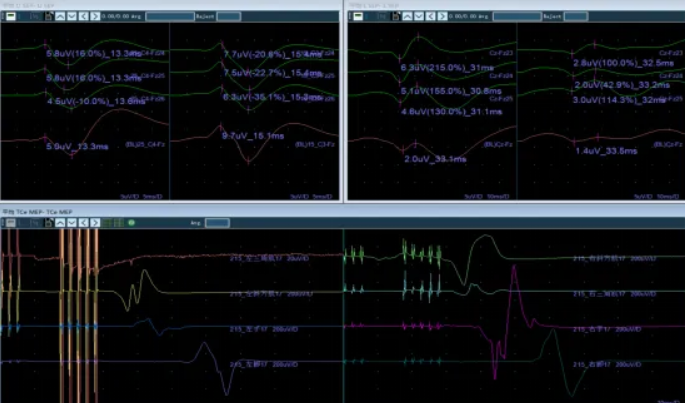
During correction and distraction at the C4 level, SEP showed no apparent change, whereas MEP waveforms disappeared. Increasing the stimulation intensity did not produce an evident recovery.
The change was immediately reported to the lead surgeon and prompted investigation. After the correction position was changed, MEP responses reappeared. Following additional fine adjustment, the amplitude did not show immediate further improvement. At the surgeon's request, all members of the surgical team stopped operative manipulation and observed for approximately 30 minutes. MEP amplitude then recovered.
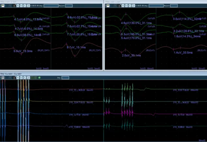
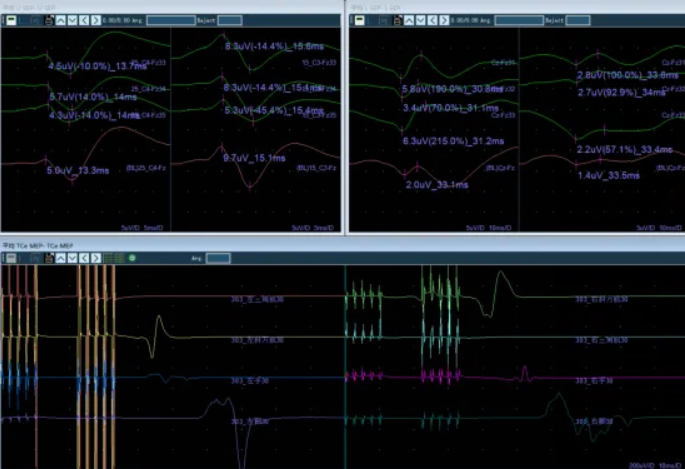
After closure, MEP and SEP were assessed again. The right upper-extremity SEP amplitude was reported to be 35% lower than the preoperative baseline, a change described in the source report as within the expected range. Lower-extremity SEP responses increased. MEP responses had essentially recovered to the preoperative level.
Congenital scoliosis correction places high demands on anesthesia because anesthetic agents can suppress neurophysiological signals and affect the accuracy of intraoperative monitoring. Preoperative communication with the anesthesiologist is essential.
TIVA with propofol and remifentanil is recommended in the source protocol. Neuromuscular blocking agents should be strictly avoided during MEP monitoring, except for limited use during induction. Although SEP is not affected by neuromuscular blocking agents, volatile anesthetics, such as sevoflurane, should be avoided because they may suppress cortical signals.
Intraoperatively, anesthetic depth must be balanced against the need for adequate sedation. Excessive anesthetic depth may decrease or abolish MEP and SEP amplitudes, whereas anesthesia that is too light may lead to patient movement and interfere with surgery.
By monitoring MEP, SEP, and Free-EMG in real time across different surgical stages, including pedicle screw placement, corrective rod installation, osteotomy closure, distraction, and compression, the clinical team can assess the effect of surgical manipulation on spinal cord and nerve-root function.
In this case, an MEP amplitude reduction of more than 75% from baseline during corrective distraction was interpreted as a warning that the spinal cord might be subject to traction or ischemic injury. The source report recommends that the surgeon immediately pause the maneuver, remove or release the corrective instrumentation as appropriate, raise mean arterial pressure to at least 80-90 mmHg, and investigate anesthetic and systemic factors, including hypothermia, anemia, and hypoxemia, to help avoid irreversible spinal cord injury.
At the end of surgery, MEP had recovered to the preoperative level. No burst, train, or sustained abnormal Free-EMG activity was reported, and SEP remained stable without significant change. The source report interpreted these findings as consistent with preservation of monitored spinal cord conduction pathways and neural function during surgery.
The procedure achieved the intended goals of deformity correction and relief of spinal cord and nerve-root compression while supporting protection of neurological function.
Congenital scoliosis is a three-dimensional spinal deformity caused by vertebral developmental abnormalities during embryogenesis, including hemivertebra, butterfly vertebra, or failure of segmentation. As a child grows, the deformity may progress rapidly. In severe cases, it can affect thoracic development, cardiopulmonary function, and body balance, or cause spinal cord compression with neurological dysfunction.
Scoliosis correction is a fundamental treatment for progressive congenital scoliosis. Its core objective is to use pedicle screw placement, osteotomy, correction, and fusion techniques to stop deformity progression while restoring spinal alignment, trunk balance, and thoracic volume to the greatest possible extent, and while protecting spinal cord, nerve-root, and vascular function.
IONM is an objective, real-time, noninvasive assessment method that can dynamically reflect spinal cord and nerve-root functional status during anesthesia. It can help determine whether internal fixation, including pedicle screws, is safely positioned; whether correction maneuvers are placing excessive traction on the spinal cord; and whether early signs of neurological injury are emerging, such as MEP amplitude reduction, SEP latency prolongation, or burst EMG activity.
In this case, real-time guidance from multimodal IONM, including SEP, MEP, and Free-EMG, supported timely adjustment of the surgical correction maneuver after an intraoperative warning. The procedure subsequently proceeded with recovery of MEP responses, while the monitoring record showed stable SEP and no significant abnormal Free-EMG activity. The case illustrates the clinical value of multimodal IONM in supporting neural protection during complex three-dimensional deformity correction and internal fixation.

Copyright © NCC Medical Co., Ltd. - Privacy policy
 EN