A case report on USEP and LSEP monitoring during aortic replacement surgery, focusing on waveform changes during bypass and arterial clamping.

A 65-year-old man was admitted with heart failure, recurrent chest tightness, palpitations, and shortness of breath. He underwent aortic replacement surgery to address a life-threatening aortic vascular lesion and reduce the risk of sudden death, as described in the source case. Intraoperative neurophysiological monitoring was used to track changes in neural pathway responses during cardiopulmonary bypass, hypothermia, aortic clamping, and left femoral artery clamping.
The case focused on serial monitoring with upper-extremity somatosensory evoked potentials (USEP) and lower-extremity somatosensory evoked potentials (LSEP). The observed changes provided time-specific information about perfusion-related physiological stress and supported continued assessment during the critical stages of the operation.
Clinical scope: This report describes one monitoring case and is intended for professional information only. SEP findings are indirect neurophysiological indicators and must be interpreted by qualified clinicians in conjunction with surgical events, hemodynamics, temperature, anesthetic conditions, and technical factors.
The primary objective of intraoperative electrophysiological monitoring in this case was to identify neurophysiological changes that could be associated with reduced perfusion during aortic replacement surgery. The source case selected SEP monitoring as the principal modality. It also notes that EEG may be used as an additional monitoring function, although EEG was not used in this case.
The source case refers to SEP monitoring in relation to brainstem function. For clinical precision, the recorded responses should be understood as indirect measures of the integrity of the monitored somatosensory pathways. Their interpretation should be integrated with the overall physiological and surgical context rather than treated as a standalone measure of cerebral function.
| Parameter | USEP | LSEP |
|---|---|---|
| Recording electrodes | C3' and C4' | Cz' |
| Reference electrode | Fz, shared | Fz, shared |
| Stimulated nerves | Left and right median nerves | Left and right posterior tibial nerves |
| Low-frequency filter | 30 Hz | 30 Hz |
| High-frequency filter | 500 Hz | 500 Hz |
| Sweep speed | 5 ms/div | 10 ms/div |
| Sensitivity | 2–5 µV | 2–5 µV |
| Stimulation rate | 2.33 Hz | 2.33 Hz |
| Stimulation intensity | 20 mA | 30 mA |
The values above reproduce the monitoring parameters reported in the source case. Actual intraoperative settings should be selected and adjusted by the monitoring team according to the patient, equipment, anesthetic conditions, and institutional protocol.
At the beginning of the procedure, baseline SEP waveforms were acquired. The recorded amplitudes and latencies were described as within the expected normal range for the case.
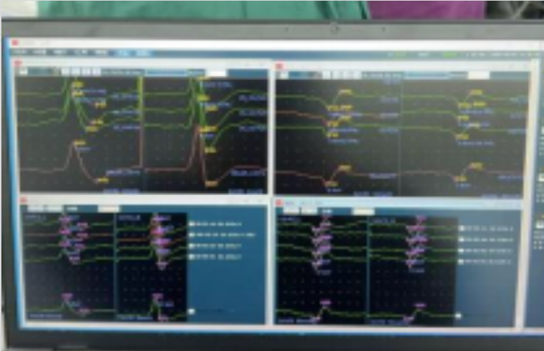
After the patient entered hypothermic cardiopulmonary bypass and the aorta and left femoral artery were clamped, the USEP latency became prolonged. The LSEP latency and amplitude decreased, and the left lower-extremity response disappeared.
These changes were interpreted in the source case as indicating reduced perfusion-related physiological stress. Because the LSEP response reflects the stimulated lower-extremity pathway, the disappearance of the left response was documented as a key side-specific monitoring change. Any comparable intraoperative change requires correlation with perfusion, hemodynamic, temperature, anesthetic, surgical, and technical information.
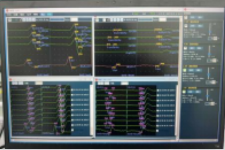
One hour later, the recorded waveforms showed no further decrease in amplitude or latency compared with the previous assessment. The source case interpreted this stability as indicating that the monitored neural function remained stable during this stage of the procedure.
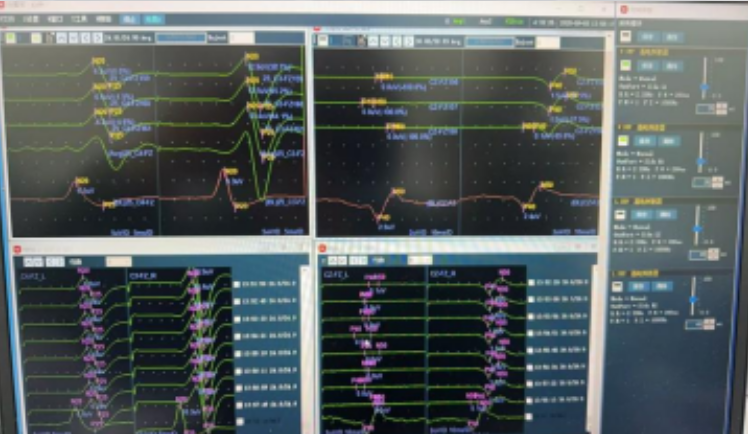
After a further 40 minutes, cardiopulmonary bypass flow was reduced in preparation for aortic unclamping. The left femoral artery remained clamped at this point.
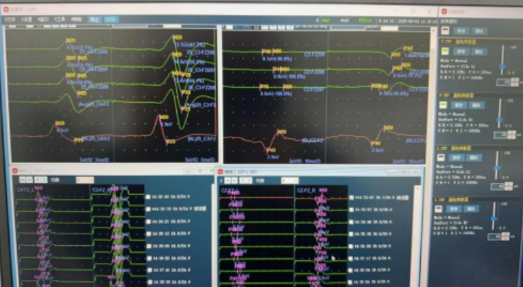
At 1 hour and 40 minutes after the start of the critical clamping and bypass sequence, aortic replacement had been completed and the left femoral artery was released. The source article does not define the timing reference more precisely. The USEP and right LSEP amplitudes and latencies returned to the reported normal range, while the left LSEP response began to recover.
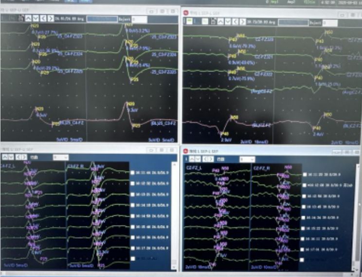
After removal of the left femoral cannula and one hour after cardiac activity had resumed, SEP amplitudes and latencies were reported as normal. The source case interpreted the final monitoring findings as showing no evident electrophysiological abnormality in the monitored responses.
The source article does not provide a detailed postoperative neurological examination, discharge status, or long-term clinical outcome. Accordingly, this report does not make a claim about postoperative neurological outcome beyond the intraoperative SEP findings described above.
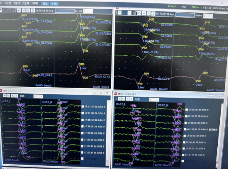
This case illustrates how serial comparison of SEP waveforms can provide time-linked neurophysiological information during major aortic surgery. Changes in latency, amplitude, and side-specific response presence occurred in temporal association with cardiopulmonary bypass, hypothermia, aortic clamping, left femoral artery clamping, and subsequent reperfusion.
The source case continued monitoring using the response after clamping as a new reference point. A persistent or progressive decline in waveform parameters should prompt immediate multidisciplinary assessment of potential causes, including:
In this context, intraoperative electrophysiological monitoring is best used as one component of a broader protective strategy. It can support early recognition of perfusion-related changes and help the surgical, anesthesia, perfusion, and monitoring teams reassess the situation during high-risk operative stages.
During aortic replacement surgery, USEP and LSEP monitoring demonstrated dynamic changes associated with cardiopulmonary bypass, hypothermia, arterial clamping, and subsequent release of the left femoral artery. The return of the monitored responses toward baseline after reperfusion provided supportive intraoperative information regarding the stability of the recorded somatosensory pathways.
This case highlights the value of continuous, time-sequenced SEP assessment during complex aortic procedures. When interpreted together with surgical and physiological data, intraoperative monitoring may help the clinical team identify possible perfusion-related risk and evaluate the effect of critical vascular interventions.

Copyright © NCC Medical Co., Ltd. - Privacy policy
 EN