Endoscopic lumbar discectomy is performed through a small working corridor close to the affected nerve root. Because the patient is under anesthesia and cannot provide reliable feedback during the procedure, the surgical team must combine anatomical visualization with appropriate intraoperative monitoring and communication.
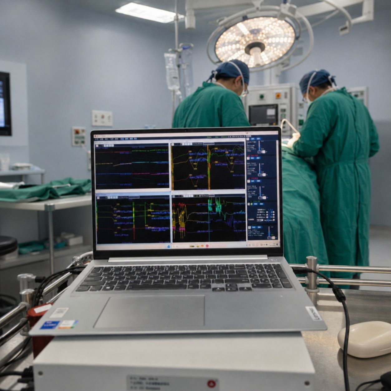
This case-based article describes the use of lower-extremity somatosensory evoked potentials (SEP), motor evoked potentials (MEP) and spontaneous electromyography (EMG) during an endoscopic lumbar discectomy. It focuses on how the monitoring modalities were incorporated into the surgical workflow, how intraoperative signal changes were interpreted, and why anesthesia and operating-room conditions matter when establishing a usable monitoring baseline.
The case involved a 76-year-old woman with lumbar spinal canal stenosis and lumbar disc herniation. Before surgery, she reported pain and weakness in both lower limbs but was still able to walk.
The surgical objective was to remove the herniated disc material and decompress the affected nerve root through an endoscopic approach. The monitoring plan combined three complementary modalities:
The combination was selected to provide information from different functional pathways rather than relying on one waveform alone.
The working space in endoscopic lumbar surgery is limited, and the nerve root may be close to the instruments and decompression field. Surgical manipulation, traction, compression or contact can affect the quality of neurophysiological signals or produce abnormal activity.
Each modality contributes different information:
Lower-extremity SEP can be recorded after stimulation of the posterior tibial nerves. In the reported case, the recording montage used a scalp recording site at Cz with Fz as the reference site, while stimulation was applied at the bilateral ankles.
SEP responses were observed from the preoperative baseline through key surgical stages. Changes in amplitude or latency should be assessed in context, with attention to technical factors, anesthesia, blood pressure, temperature, electrode contact and surgical events. A waveform change alone should not be interpreted as a diagnosis of neurological injury.
MEP responses were recorded from muscles supplied by the lower-limb nerve roots, including the quadriceps, tibialis anterior, gastrocnemius and abductor hallucis. Transcranial stimulation was applied over the motor cortex.
MEP can be sensitive to anesthetic management and neuromuscular blockade. In this case, the initial MEP amplitude was limited while muscle relaxation had not fully resolved. A later measurement after the effect of muscle relaxation had decreased produced a stronger response, illustrating why the timing and conditions of baseline acquisition are important.
Spontaneous EMG was recorded from lower-limb muscles. During endoscopic exploration and disc removal, bursts of EMG activity were observed in the tibialis anterior and gastrocnemius muscles.
Such activity may indicate mechanical irritation or stimulation of a nerve root. It should prompt the monitoring and surgical teams to review the current surgical maneuver, equipment conditions and possible sources of interference. The appropriate response should follow the team’s established alarm and communication protocol.
Before monitoring begins, the team should review the patient’s preoperative symptoms and any available neurological or electrodiagnostic information. These findings provide clinical context for interpreting intraoperative responses.
The monitoring team should also confirm:
A baseline should be recorded under conditions that are clearly documented. If the physiological or anesthetic conditions change substantially, the team should consider how those changes affect comparisons with the baseline.
During puncture and endoscope placement, the case report described stable resting EMG and recognizable SEP responses. MEP responses were relatively limited at the early stage because the effect of muscle relaxation had not completely resolved.
During endoscopic exploration, the surgical team identified the relevant anatomical structures and proceeded toward decompression. A decrease in the right-sided SEP response was observed during part of the exploration. At the same time, burst EMG activity appeared in a lower-limb muscle during disc manipulation.
These observations illustrate the practical role of multimodal monitoring: a change in one modality can be considered alongside the other waveforms and the exact surgical maneuver being performed.
During removal of the herniated disc material, changes in the lower-limb SEP responses and burst EMG activity were observed. The monitoring team followed the waveforms as the surgical maneuver progressed and communicated relevant changes to the surgical team.
For a clinical article, it is important to describe this process without implying that a single waveform change automatically confirms injury. Signal interpretation depends on the quality of the recording, the baseline, anesthetic and physiological conditions, the surgical event and the response after the maneuver is paused or adjusted.
At the end of the procedure, the team repeated SEP and MEP monitoring. The case report described a recovery trend in the right-sided SEP response compared with the intraoperative decrease, while the MEP response did not show a substantial deterioration from the exploration stage.
The final interpretation was made together with the surgical events and the overall waveform trend. Intraoperative monitoring can support communication and decision-making, but it should not be presented as an independent guarantee of postoperative outcome.
The quality of SEP, MEP and EMG monitoring is influenced by more than the monitoring system itself. The case highlights several factors that should be considered before and during endoscopic lumbar surgery.
Neuromuscular blocking medication can reduce or suppress EMG and MEP responses. The monitoring team and anesthesia team should therefore discuss the planned medication and timing before baseline acquisition. The protocol should distinguish the requirements for airway management from the requirements for reliable neurophysiological monitoring.
Anesthetic agents may affect evoked potentials. Stable anesthesia helps the team distinguish physiological or medication-related changes from changes associated with a surgical maneuver. The selected anesthetic plan should be documented and interpreted by qualified anesthesia and monitoring professionals.
Changes in blood pressure and body temperature can influence neurophysiological recordings. Maintaining stable physiological conditions supports more consistent interpretation, although it does not eliminate all sources of variability.
Electrosurgical equipment and other operating-room devices may introduce artifacts. When a waveform changes unexpectedly, the team should consider electrode contact, cables, stimulation settings, equipment interference and physiological conditions before attributing the change to surgical manipulation.
This case illustrates several practical points for IONM in spine surgery:
Endoscopic lumbar discectomy places surgical instruments close to the nerve root in a setting where the patient cannot provide real-time neurological feedback. A monitoring plan that combines SEP, MEP and spontaneous EMG can provide complementary information during access, exploration, disc removal, decompression and closure.
The case also shows why effective IONM depends on more than waveform acquisition. Baseline recording, anesthesia coordination, physiological stability, artifact control and communication with the surgical team all contribute to a meaningful intraoperative workflow.
For hospitals evaluating an IONM solution for spine surgery, the key question is not simply whether a system supports multiple modalities. The evaluation should also consider how the system, electrodes, stimulation accessories, reporting functions, training and technical support fit the hospital’s actual surgical workflow.

Copyright © NCC Medical Co., Ltd. - Privacy policy
 EN
EN